A scanning probe microscope tip coated with a wax layer and its preparation method and application

A scanning probe and microscope technology, used in scanning probe microscopy, scanning probe technology, instruments, etc., can solve the problems of difficult removal of large pollutants, easy damage to modified layers, and damage to the tip of the needle tip. The realization method is simple and easy to operate, the surface of the needle tip is clean, and the cost is low.

Active Publication Date: 2020-02-21
THE NAT CENT FOR NANOSCI & TECH NCNST OF CHINA
View PDF2 Cites 0 Cited by
  • Summary
  • Abstract
  • Description
  • Claims
  • Application Information

AI Technical Summary

Problems solved by technology

However, these methods have many deficiencies in removing pollutants on the surface of the AFM needle tip: first, the above methods are only suitable for the removal of thin layers of organic pollutants, and it is difficult to remove large pollutants; The harsh processing environment of the above method is easy to be damaged and fall off; secondly, during the cleaning process, the tip of the needle tip is very easy to be damaged due to mechanical collisions and other reasons
For these reasons, in practical applications, contaminated needle tips are discarded in most cases

Method used


Image

  • A scanning probe microscope tip coated with a wax layer and its preparation method and application
  • A scanning probe microscope tip coated with a wax layer and its preparation method and application
  • A scanning probe microscope tip coated with a wax layer and its preparation method and application

Examples


Embodiment 1

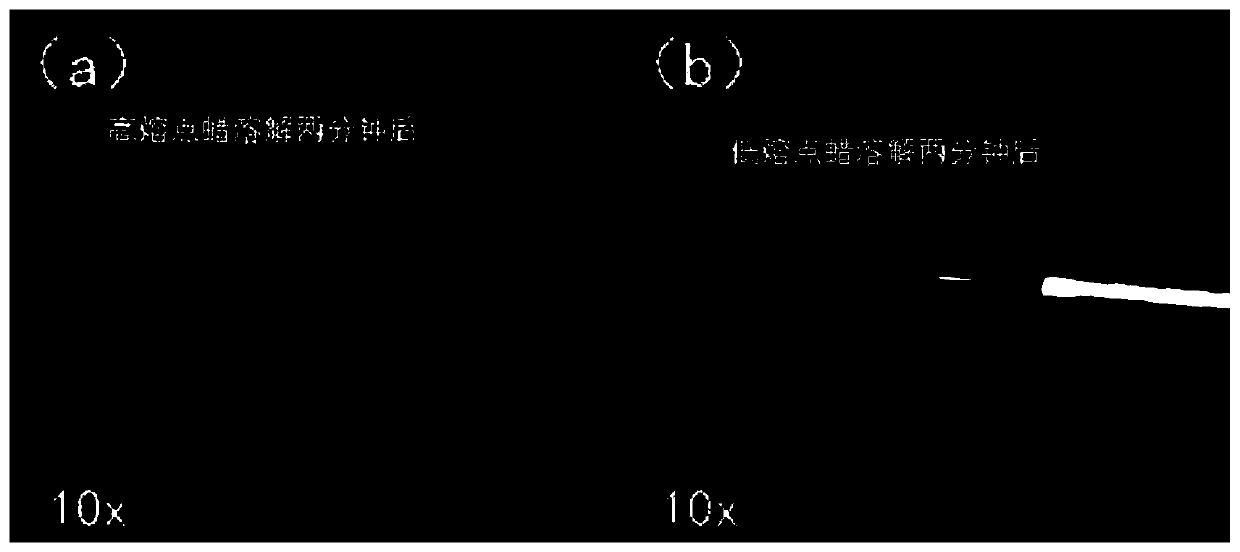
[0031] Example 1 High / Low Melting Point Wax Removal Comparative Experiment

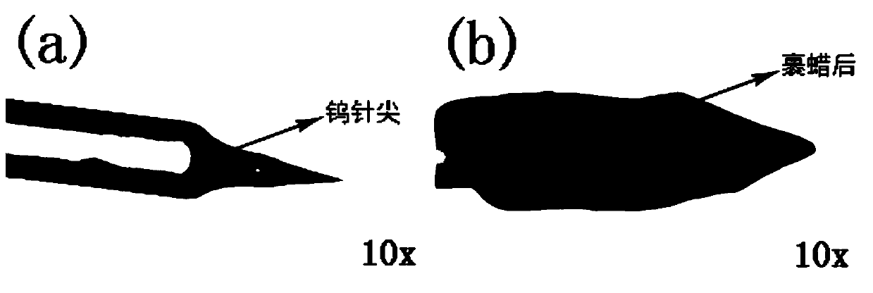
[0032] Put the low melting point paraffin into a clean container, heat the container until the solid paraffin is completely melted to the liquid paraffin, and the heating temperature is controlled near the melting point of the paraffin. The tungsten probe tip obtained by chemical etching ( figure 1 a) Submerge vertically below the liquid paraffin surface so that the needle tip is just submerged in the paraffin liquid surface. Immediately pull the tungsten probe out of the paraffin, and cool at room temperature to solidify the paraffin. The tungsten probe tip is pulled out from the liquid paraffin for a moment, and the liquid paraffin dipped on the surface of the tip solidifies immediately to form a solid wax protective layer on the surface of the tungsten tip (such as figure 1 shown in b).

[0033] Fix the tip of the above-mentioned wax-coated tungsten needle so that the tip part is submerged in th...

Embodiment 2

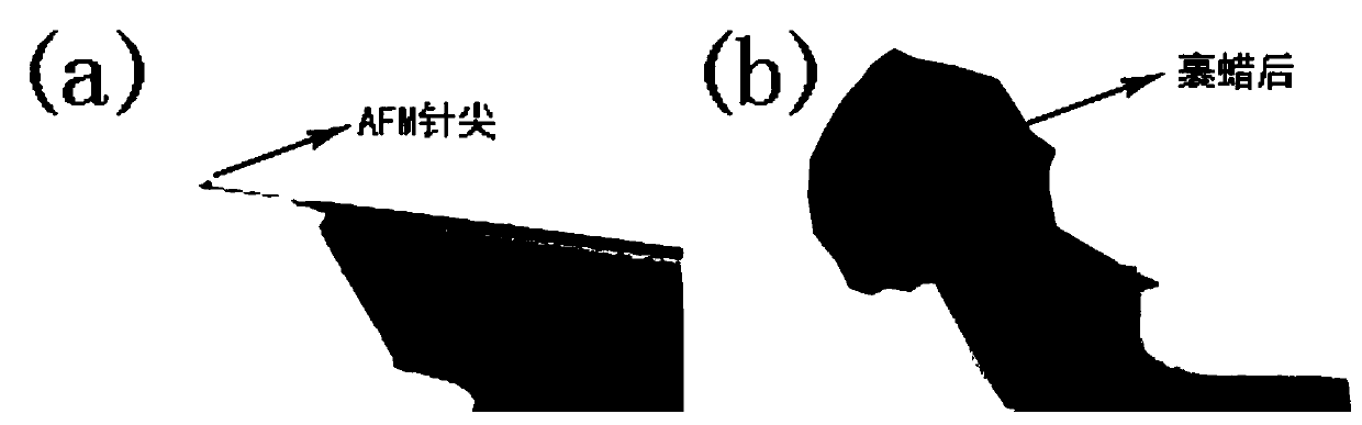
[0036] Put the low melting point paraffin into a clean container, heat the container until the solid paraffin is completely melted to the liquid paraffin, and the heating temperature is controlled near the melting point of the paraffin. The AFM probe ( figure 2a) The cantilever part is submerged vertically below the liquid paraffin surface, so that the needle tip is just submerged in the paraffin liquid surface. Immediately extract the AFM probe cantilever from the paraffin, and cool at room temperature until the paraffin solidifies. The AFM probe cantilever is pulled out of the liquid paraffin for a moment, and the liquid paraffin dipped on its surface solidifies immediately to form a solid wax protective layer on the surface of the cantilever ( figure 2 b).

[0037] The wax protective layer on the surface of the AFM probe cantilever covered with wax can be removed by infrared baking and melting to remove the wax: place the AFM probe covered with wax vertically on the bas...

Embodiment 3

[0039] Put the low melting point paraffin into a clean container, heat the container until the solid paraffin is completely melted to the liquid paraffin, and the heating temperature is controlled near the melting point of the paraffin. Submerge the cantilever part of the AFM probe vertically below the liquid paraffin surface, so that the needle tip is just submerged in the paraffin liquid surface. Immediately extract the AFM probe cantilever from the paraffin, and cool at room temperature until the paraffin solidifies. The AFM probe cantilever is pulled out from the liquid paraffin for a moment, and the liquid paraffin dipped on its surface solidifies immediately to form a solid wax protective layer on the surface of the cantilever (such as figure 2 shown in b).

[0040] The wax protective layer on the surface of the wax-coated AFM probe cantilever can be removed by dissolving the wax in an organic solvent: immerse the wax-coated AFM probe cantilever into the organic solven...


PUM

PropertyMeasurementUnit
thicknessaaaaaaaaaa
lengthaaaaaaaaaa
melting pointaaaaaaaaaa

Abstract

The invention relates to a waxed probe tip of a scanning probe microscope and a preparation method and application thereof. The tip part of a probe tip is waxed so as to keep the probe tip clean, such that the protected probe tip material keeps the probe tip at an original state and is prevented from being polluted by all kinds of foreign impurities. According to the invention, the method is simple and and can better protect the probe tip. The de-waxing of the waxed probe tip material is easy to operate with no waxes left and no damage to the probe tip structure, and saves excellent imaging and measuring functions of the probe tip.

Description

technical field [0001] The invention relates to a scanning probe microscope tip coated with a wax layer, a preparation method and application thereof. Background technique [0002] Scanning probe microscopy (SPM) is a general term for scanning tunneling microscopy (STM) and various probe microscopy techniques developed on its basis. Including atomic force microscopy (AFM) technology, laser force microscopy (LFM) technology, magnetic force microscopy (MFM) technology, etc. Among them, atomic force microscopy (AFM) technology is the most widely used technology in the scanning probe microscopy technology family. It is widely used in materials science, semiconductor research and development, microbiology and other fields because of its excellent detection ability of surface morphology characterization. It has greatly accelerated the development of related fields and has become a basic tool for research in the field of nanoscience today. [0003] The probe tip of the atomic for...

Claims


Application Information

Patent Timeline
Patent Type & Authority Patents(China)
IPC IPC(8): G01Q60/04G01Q60/38G01Q60/16
CPCG01Q60/04G01Q60/16G01Q60/38
Inventor 徐建勋卢兴徐乐乐黄威
Owner THE NAT CENT FOR NANOSCI & TECH NCNST OF CHINA
Why Eureka
Try Eureka