Method for performing Hpx protein induction and maintaining selective polarization of microglial cells, and applications thereof
A technology of microglia and protein, applied in the fields of bioengineering and medicine, can solve the problems that have not yet been reported on the polarization of Hpx protein
- Summary
- Abstract
- Description
- Claims
- Application Information
AI Technical Summary
Problems solved by technology
Method used
Image
Examples
Embodiment 1
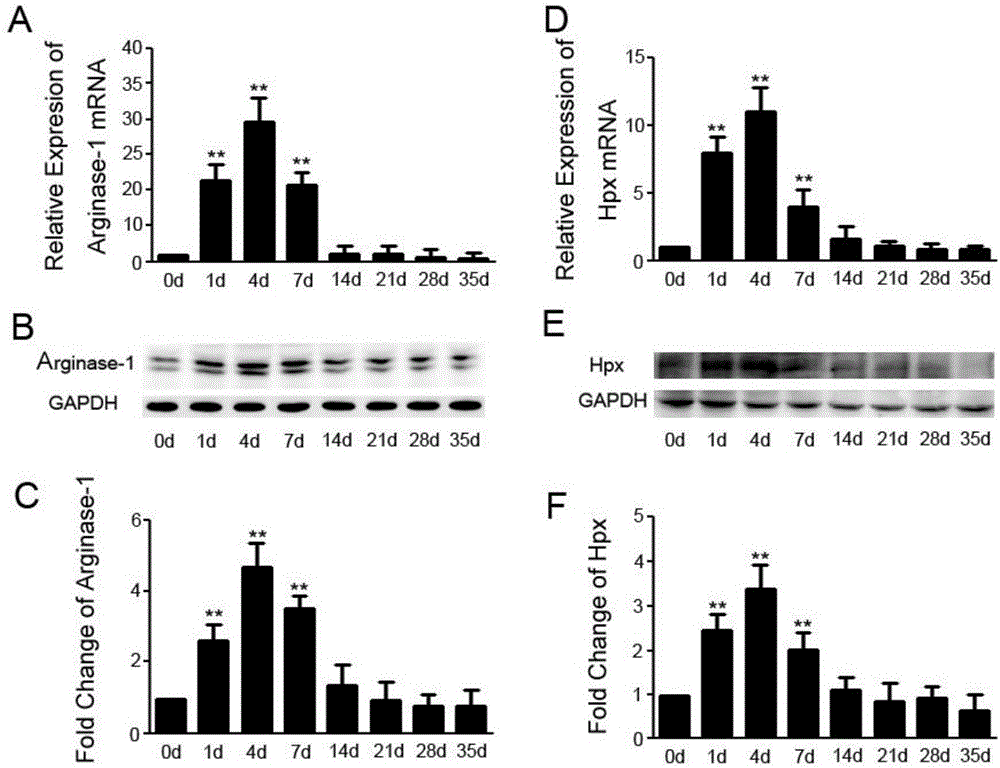
[0051] Example 1: Modeling of spinal cord injury, detection of correlation between Hpx and microglial cell polarization markers.
[0052] 1. Spinal cord injury modeling
[0053] After anesthetized by intraperitoneal injection of 4% chloral hydrate, the mice were placed prone on the foam board after skin preparation. The mouse spine was passively flexed, and the most obvious kyphosis of the thoracolumbar segment was calculated to the head side for 1-2 stages, and a longitudinal surgical incision was taken. Cut the skin, separate the bilateral musculature, reveal the spinous process and bilateral lamina, and locate the lamina stage. At chest 9-10, use a rongeur to bite off the 9th lamina of the chest to fully expose the spinal cord tissue. The spinal cord tissue can be seen to be bright white, and the longitudinal artery can be seen in the posterior center. If it is a control operation of laminectomy, the surgical incision can be sutured at this time; if it is a spinal cord c...
Embodiment 2
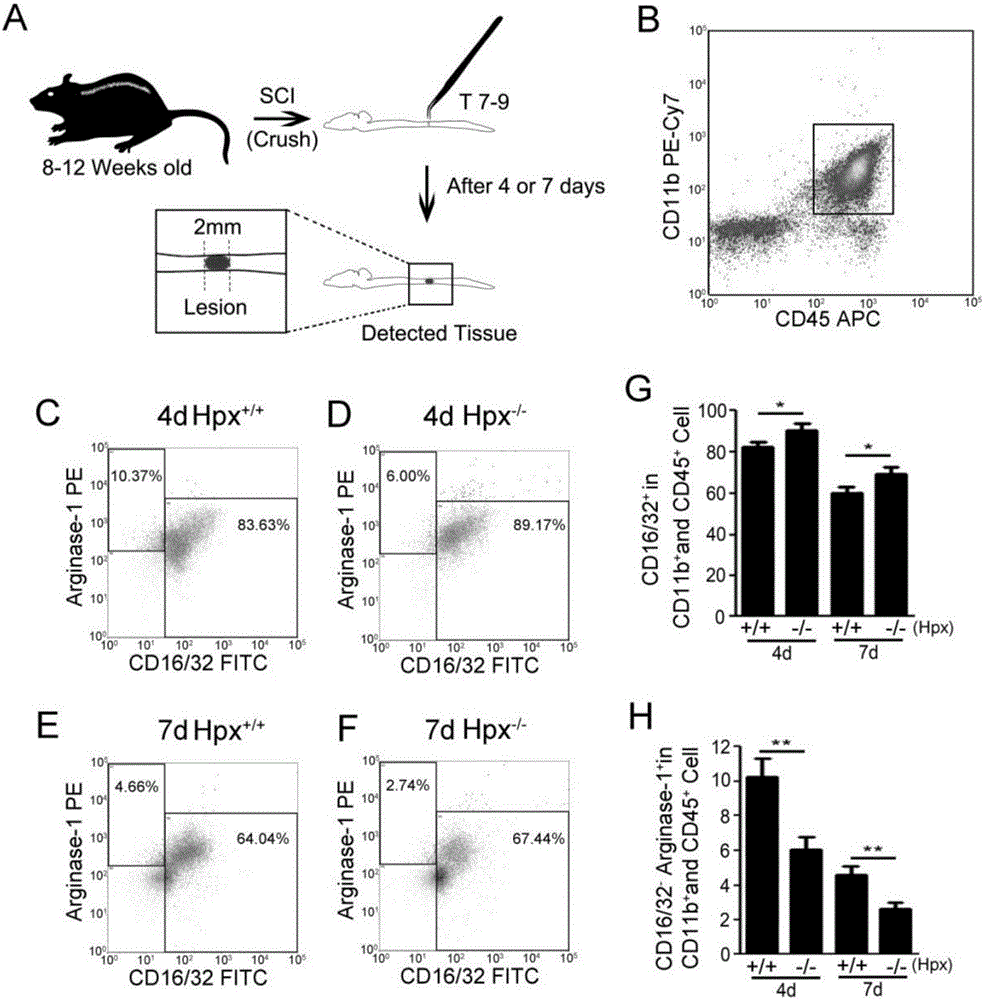
[0069] Example 2: Flow Cytometry Analysis
[0070] The microglial cells in the injured spinal cord of Hpx knockout mice were sorted by flow cytometry, and the polarization ratio was detected.
[0071] A. Collection of monocytes
[0072] The mice that have been modeled for a certain number of days were anesthetized by intraperitoneal injection of 4% chloral hydrate. After the anesthesia was complete, they were fixed on the foam board in a supine position, and the upper and lower limbs were pulled longitudinally as much as possible to fully stretch the spine structure. Make a "U"-shaped incision on the chest, cut the skin, cut the ribs, expose the heart, cut the right atrial appendage, and then puncture the left ventricle with an injection needle. First, use 1×PBS solution for continuous injection, about 40ml. Then the spinal cord tissue (including 1 mm above and below the spinal cord injury) was surgically removed under a microscope and placed in dissecting fluid at 4°C. The ...
Embodiment 3
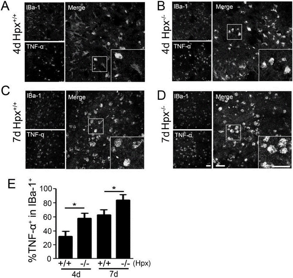
[0076] Embodiment 3: histochemical experiment
[0077] Using immunohistochemical method, the expression level of M1 polarization marker molecule TNF-a in microglial cells of Hpx knockout mice with spinal cord injury was analyzed to further verify the regulation of Hpx on the polarization of microglial cells.
[0078] A. Histochemical staining steps
[0079] After placing the frozen sections in a 37°C oven for 1 hour, follow the steps below: wash the slices twice for 5 minutes with PBS; incubate overnight at 4°C with the anti-TNF-a primary antibody diluted in PBS; let the slices stand at room temperature for 40 minutes; wash with PBS at room temperature Slices, 5 minutes each time, three times in total; add secondary antibody, incubate at room temperature for 70 minutes; wash the slides with normal temperature PBS, 5 minutes each time, three times in total; then incubate with 1:1000 Hoechst for 7 minutes; wash with normal temperature PBS Films, 5 minutes each time, three times...
PUM
 Abstract
Description
Claims
Application Information
 - R&D
- Intellectual Property
- Life Sciences
- Materials
- Tech Scout
- Unparalleled Data Quality
- Higher Quality Content
- 60% Fewer Hallucinations
Browse by: Latest US Patents, China's latest patents, Technical Efficacy Thesaurus, Application Domain, Technology Topic, Popular Technical Reports.
© 2025 PatSnap. All rights reserved.Legal|Privacy policy|Modern Slavery Act Transparency Statement|Sitemap|About US| Contact US: help@patsnap.com


