Piglet small intestine epithelial cell classification and separation method

A technology for intestinal epithelial cells and epithelial cells is applied in the field of piglet intestinal epithelial cell fractionation, which can solve the problems of inability to realize high-throughput research on physiological processes and metabolic changes, and achieve the effects of low cost and simple operation.

Active Publication Date: 2017-05-17
HUNAN NORMAL UNIVERSITY
View PDF2 Cites 6 Cited by
  • Summary
  • Abstract
  • Description
  • Claims
  • Application Information

AI Technical Summary

Problems solved by technology

The only few in vivo studies mainly use rodents such as mice and rats as models, and use techniques such as immunohistochemistry to study changes in a small amount of gene expression or protein synthesis, which cannot achieve relevant physiological processes and metabolic changes in the process of intestinal cell differentiation high-throughput research
Moreover, due to the short gestation period, the intestinal development pattern of rodents is quite different from that of humans, so it is very limited to use rodents as a model to study the differentiation process and developmental changes of human intestinal epithelial cells

Method used


Image

  • Piglet small intestine epithelial cell classification and separation method
  • Piglet small intestine epithelial cell classification and separation method
  • Piglet small intestine epithelial cell classification and separation method

Examples


Embodiment 1

[0025] The method for fractionating piglet intestinal epithelial cells comprises the following steps:

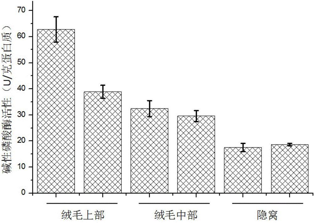
[0026] (1) Six 0-day-old suckling piglets were selected. After slaughtering, the small intestines of piglets with a length of 60 cm were cut, and 100 mL of preheated oxygenated phosphate buffer solution at 37 ° C was perfused into the intestinal cavity, and both ends were clamped with hemostatic forceps. Incubate in a water bath shaker at ℃ for 30 minutes at a shaking speed of 70 rpm;

[0027] Among them, the phosphate buffer solution is composed of: sodium chloride 150mM, sodium dihydrogen phosphate 4mM, disodium hydrogen phosphate 4mM, phenylmethylsulfonyl fluoride 0.1mM, dithiothreitol 0.4mM, pH 7.4.

[0028] (2) Discard the phosphate buffer, perfuse 100mL of cell separation solution, and continue to incubate in a water bath shaker at 37°C for 2.5 hours at a shaking speed of 70 rpm. Minutes, 120 minutes and 150 minutes to collect cell separation fluid and perfuse new cel...

Embodiment 2

[0033] The method for fractionating piglet intestinal epithelial cells comprises the following steps:

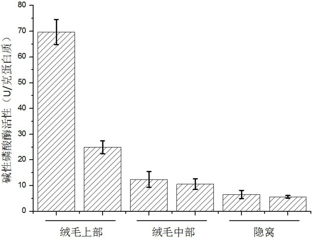
[0034] (1) Six 25-day-old weaned piglets were selected. After slaughtering, the piglets’ small intestines with a length of 80 cm were cut, and 110 mL of preheated oxygenated phosphate buffer solution at 37 ° C was perfused into the intestinal cavity, and both ends were clamped with hemostatic forceps. Incubate in a water bath shaker at ℃ for 30 minutes at a shaking speed of 70 rpm;

[0035] Among them, the phosphate buffer solution is composed of: sodium chloride 142mM, sodium dihydrogen phosphate 5mM, disodium hydrogen phosphate 5mM, phenylmethylsulfonyl fluoride 0.2mM, dithiothreitol 0.5mM, pH 7.4.

[0036] (2) Discard the phosphate buffer, perfuse 110mL of cell separation solution, and continue to incubate in a water bath shaker at 37°C for 2.5 hours at a shaking speed of 70 rpm. Minutes, 115 minutes and 150 minutes to collect cell separation fluid and perfuse new cell s...

Embodiment 3

[0041] The method for fractionating piglet intestinal epithelial cells comprises the following steps:

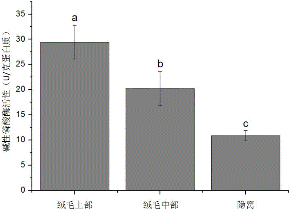
[0042] (1) Select 6 70-day-old piglets, cut the small intestine section with a length of 90cm after slaughtering, perfuse 150mL of preheated oxygenated phosphate buffer solution at 37°C in the intestinal cavity, clamp both ends with hemostatic forceps, and incubate at 37°C Incubate in a water bath shaker for 30 minutes at a shaking speed of 70 rpm;

[0043] Among them, the phosphate buffer solution is composed of: sodium chloride 135mM, sodium dihydrogen phosphate 6mM, disodium hydrogen phosphate 6mM, phenylmethylsulfonyl fluoride 0.3mM, dithiothreitol 0.6mM.

[0044] (2) Discard the phosphate buffer, perfuse 150mL of the cell separation solution, and continue to incubate in a water bath shaker at 37°C for 2.5 hours at a shaking speed of 70 rpm, and collect the cells at 40 minutes, 90 minutes and 150 minutes after incubation. Separation fluid and perfuse new cell separation f...


PUM


Abstract

The invention discloses a piglet small intestine epithelial cell classification and separation method, which includes the following steps: (1) a small intestine section which is 60cm to 90cm long is adopted, 100mL to 150mL of 37 DEG C preheated and oxygenated phosphate buffer is injected into the enteric cavity, both ends are clipped by hemostatic forceps, the small intestine section is then incubated in a 37 DEG C water bath oscillator for 30 minutes, and the speed of oscillation is 70 rotations per minute; (2) the phosphate buffer is removed, 100mL to 150mL of cell separation solution is injected, the small intestine section continues to be incubated in the 37 DEG C water bath oscillator for 2.5 hours, the speed of oscillation is 70 rotations per minute, and the cell separation solution is collected respectively at different time points and new cell separation solution is injected; (3) the collected cell separation solution is centrifuged at 400g under 4 DEG C for ten minutes, the obtained precipitate is intestinal epithelial cells, and the cells which are collected according to the sequence of incubation times are cells of different parts from the villus tip to the crypt bottom in sequence along a recess-villus axis. The cells separated by the invention provide a novel technical method for an in-vivo study on the small intestine epithelial cell differentiation mechanism, the absorption and metabolism of nutrition and the development of drugs for related diseases.

Description

technical field [0001] The invention belongs to the field of biotechnology, and in particular relates to a method for fractionating piglet small intestinal epithelial cells. Background technique [0002] The intestinal epithelium is a selective barrier for direct contact between the intestinal tract and the external environment, and plays a vital role in the digestion and absorption of nutrients and in resisting the invasion of pathogens such as bacteria. The epithelial cells of the small intestine are in a process of continuous renewal. The differentiated mature villi top epithelial cells continue to apoptotic and discharge into the intestinal cavity, while the stem cells located in the crypts continue to divide to produce new cells, and migrate along the crypt-villus axis to supplement apoptosis. The epithelial cells maintain the stability of the intestinal epithelium, and the relative stability of the intestinal epithelium is the basis for maintaining its function. The k...

Claims


Application Information

Patent Timeline
Patent Type & Authority Applications(China)
IPC IPC(8): C12N5/071
CPCC12N5/0625C12N5/0679C12N2509/00
Inventor 杨焕胜印遇龙李建中
Owner HUNAN NORMAL UNIVERSITY
Why Eureka
Try Eureka