
Double-cavity uterus contrast water through pipe

A technology for water tube and uterus, which is used in catheters, balloon catheters, medical science, etc., can solve the problems of high price of imaging tubes, rupture of balloons, complicated shapes, etc., so as to avoid the risk of radiation, slow down the expansion speed, and avoid reverse flow. Effect

Pending Publication Date: 2017-05-31
THE 309TH HOSPITAL OF CHINESE PEOPLES LIBERATION ARMY
View PDF6 Cites 1 Cited by
  • Summary
  • Abstract
  • Description
  • Claims
  • Application Information

AI Technical Summary

Problems solved by technology

The existing problems are: for patients with a small uterine cavity, the head end of the contrast tube will be bent due to the increase of pressure during the injection, resulting in blocking the interstitial part of the fallopian tube, so that the ipsilateral fallopian tube will not be developed and cause false positives; at the same time, because there is no control valve , Doctors cannot leave the operating room after injecting drugs, which virtually increases the risk of radiation exposure to doctors and patients
[0004] At present, the double-balloon imaging tubes on the market are all three-lumen tubes. The drug injection and double-balloon are separate lumens, which are made of hard plastic materials. This type of imaging tube is expensive and complicated in shape. , the doctor may confuse the medicine into the balloon and cause the balloon to rupture, especially if the intrauterine balloon ruptures, it must be replaced with a new tube

Method used


Image

  • Double-cavity uterus contrast water through pipe
  • Double-cavity uterus contrast water through pipe
  • Double-cavity uterus contrast water through pipe

Examples


Embodiment Construction

[0032] The present invention will be further described in detail below in conjunction with the accompanying drawings and specific embodiments.

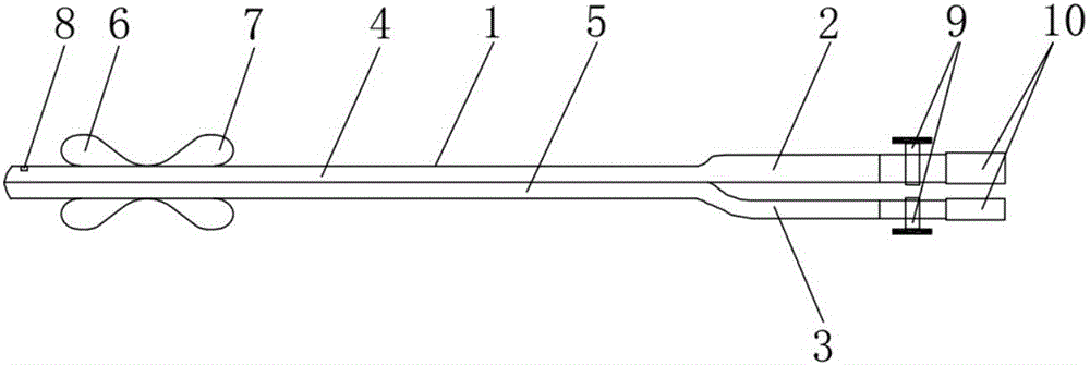
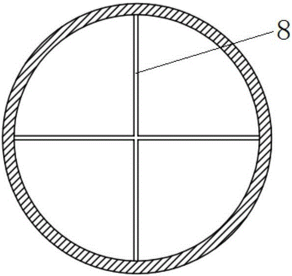
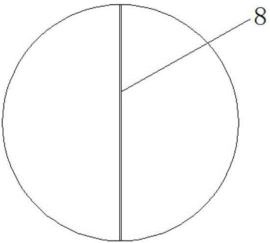
[0033] Such as figure 1 As shown, the present invention provides a double-chamber hysterography water pipe, comprising a main pipe 1 and a liquid injection connecting pipe 2 and an inflation connecting pipe 3 arranged at the rear end of the main pipe, and a mutually isolated main chamber channel 4 and a ball The sac cavity channel 5, wherein the main cavity channel 4 communicates with the injection connecting tube 2, the balloon cavity channel 5 communicates with the inflation connecting tube 3, and the front end of the main tube 1 is provided with a balloon that communicates with the balloon cavity channel 5 A liquid outlet 8 with an anti-backflow opening is formed on the main cavity channel 4 at the front end of the main tube 1. When the injection pressure in the main cavity channel 4 is greater than the pressure in the uterine cavi...


PUM


Abstract

The invention discloses a double-cavity uterus contrast water through pipe, and belongs to the technical field of medical apparatuses and instruments. The double-cavity uterus contrast water through pipe comprises a main pipe, a liquid injection connecting pipe and an air inflation connecting pipe, wherein the liquid injection connecting pipe and the air inflation connecting pipe are arranged at the rear end of the main pipe, the main pipe is provided with a main cavity passageway and a sacculus cavity passageway which are isolated from each other, the main cavity passageway is in communication with the liquid injection connecting pipe, the sacculus cavity passageway is in communication with the air inflation connecting pipe, and the front end of the main pipe is provided with a sacculus which is in communication with the sacculus cavity passageway; a liquid outlet which is provided with a backflow preventing open hole is molded on the main cavity passageway which is located at the front end of the main pipe, when the liquid injection pressure in the main cavity passageway is larger than uterine cavity pressure, the backflow preventing open hole is opened, and the injection liquid in the main cavity passageway flows out. According to the double-cavity uterus contrast water through pipe, the backflow preventing open hole is arranged at the front end of the main cavity passageway, only when the pressure injected into the main cavity passageway is larger than the uterine cavity pressure, the backflow preventing open hole can be opened so as to push the injection liquid into the uterine cavity; after drug injecting is stopped, the backflow preventing open hole is at a closed state, and thus the backflow of drug in the uterine cavity is avoided.

Description

technical field [0001] The invention belongs to the technical field of medical devices, in particular to a double-chamber hysterography water conduit. Background technique [0002] In recent years, the infertility of women of childbearing age has been increasing year by year, among which tubal obstruction is the main cause, especially in secondary infertility, the proportion can be as high as 66.67%. There are many ways to check the patency of fallopian tubes, including imaging examination, tubal fluid and salpingoscopic laparoscopy, and imaging examinations include hysterosalpingography, hysterosalpingography and so on. Tubal fluid is simple and easy to do, but there are large blindness and inaccuracy, and it is currently used as a primary screening method. Laparoscopic tubal fluid examination under direct vision is currently considered the most reliable examination method, but due to the invasiveness and high price of laparoscopy, it is more inclined to be used as a treat...

Claims


Application Information

Patent Timeline
Patent Type & Authority Applications(China)
IPC IPC(8): A61M25/10A61B17/42A61B5/00A61M29/04
CPCA61M25/0026A61M25/0043A61M25/1011A61M25/1018A61M29/02A61B5/00A61B5/4325A61B5/6853A61B5/6875A61B17/42A61B2017/4233A61M2210/1425A61M2210/1433A61M2025/1013
Inventor 杨艳敖国昆董莘
Owner THE 309TH HOSPITAL OF CHINESE PEOPLES LIBERATION ARMY
Learn More