Method and Apparatus for Tissue Ablation

a tissue and ablation technology, applied in the field of medical equipment and procedures, can solve the problems of high risk of complications, bleeding and perforation, and difficult removal of flat sessile polyps, and achieve the effect of preventing condensation of said vapor

Active Publication Date: 2013-01-03
SANTA ANNA TECH LLC
View PDF5 Cites 114 Cited by
  • Summary
  • Abstract
  • Description
  • Claims
  • Application Information

AI Technical Summary

Benefits of technology

[0034]In one embodiment, the tissue ablation system further comprises a metal frame within said catheter, wherein said metal frame is in thermal contact with said heating chamber and conducts heat to said catheter lumen, thereby preventing condensation of said va

Problems solved by technology

While most polyps are detected on colonoscopy and easily removed using a snare, flat sessile polyps are hard to remove using the snare technique and carry a high risk of complications, such as bleeding and perforation.
The presence of submucosal uterine fibroids and a large uterus size have been shown to decrease the efficacy of standard endometrial ablation.
Conventional techniques have inherent limitations, however, and have not found widespread clinical applications.
This ablation mechanism is operator dependent, cumbersome and time consuming.
Second, because the target tissue is moving due to patient movement, respiration movement, normal peristalsis and vascular pulsations, the depth of ablation of the target tissue is inconsistent and results in a non-uniform ablation.
Superficial ablation results in incomplete ablation with residual neoplastic tissue left behind.
Deeper ablation results in complications such as bleeding, stricture formation and perforation.
All of these limitations and complications have been reported with conventional devices.
However, the rigid electrode does not accommodate for variations in esophageal size and is ineffective in ablating lesions within a tortuous esophagus, proximal esophageal lesions as an esophagus narrows toward the top, and lesions in the esophagus at the gastroesophageal junction due to changes in the esophageal diameter.
Nodular disease in Barrett's esophagus also cannot be treated using the rigid bipolar RF electrode.
Due to its size and rigidity, the electrode cannot be passed through the scope.
In addition, sticking of sloughed tissue to the electrode impedes delivery of radiofrequency energy resulting in incomplete ablation.
The tissue is then exposed to a selected wavelength of light which activates the photo-sensitizer and results in tissue destruction.
PDT is associated with complications such as stricture formation and photo-sensitivity which has limited its use to the most advanced stages of the disease.
In addition, patchy uptake of the photo

Method used


Image

  • Method and Apparatus for Tissue Ablation
  • Method and Apparatus for Tissue Ablation
  • Method and Apparatus for Tissue Ablation

Examples


Embodiment Construction

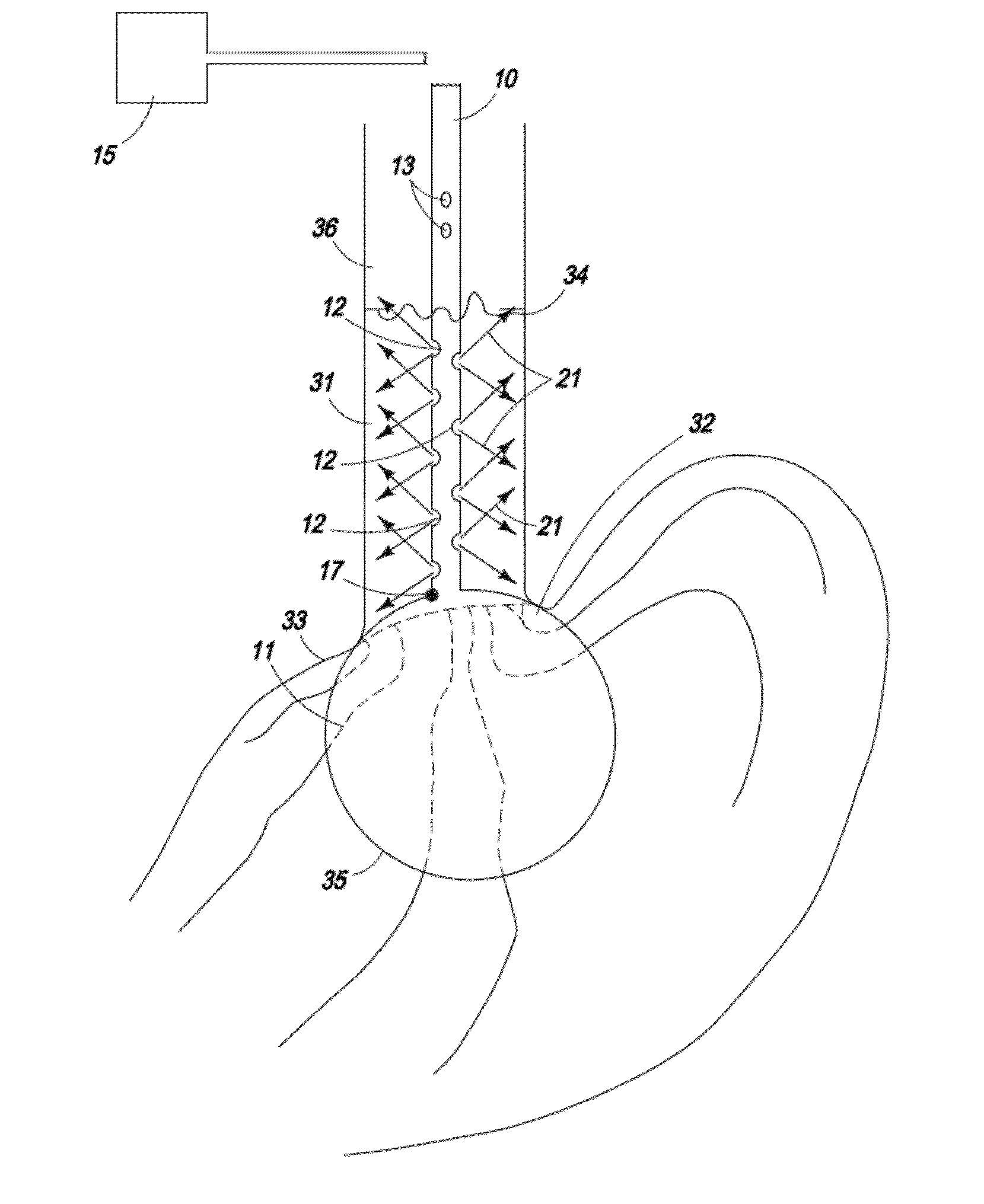
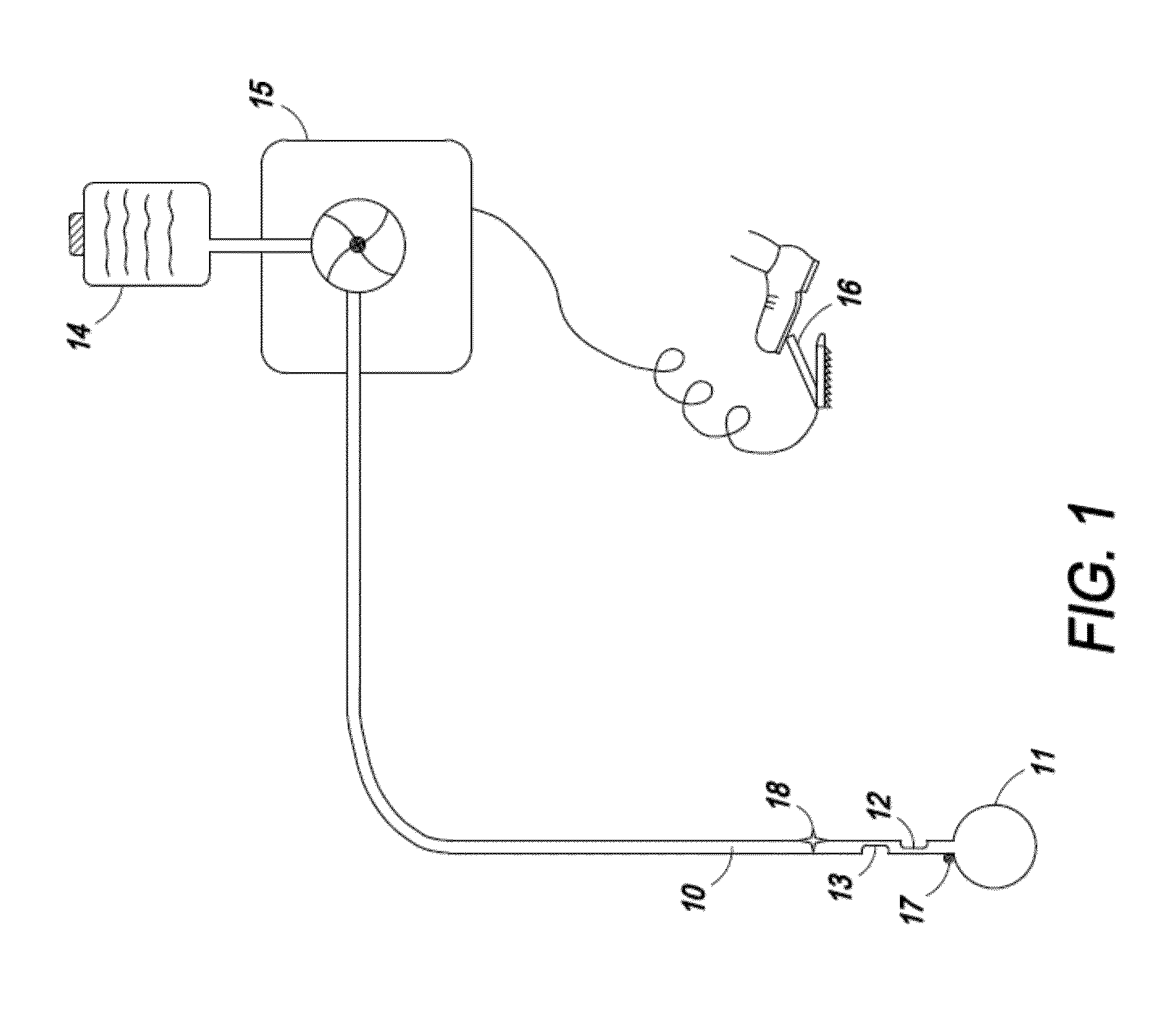
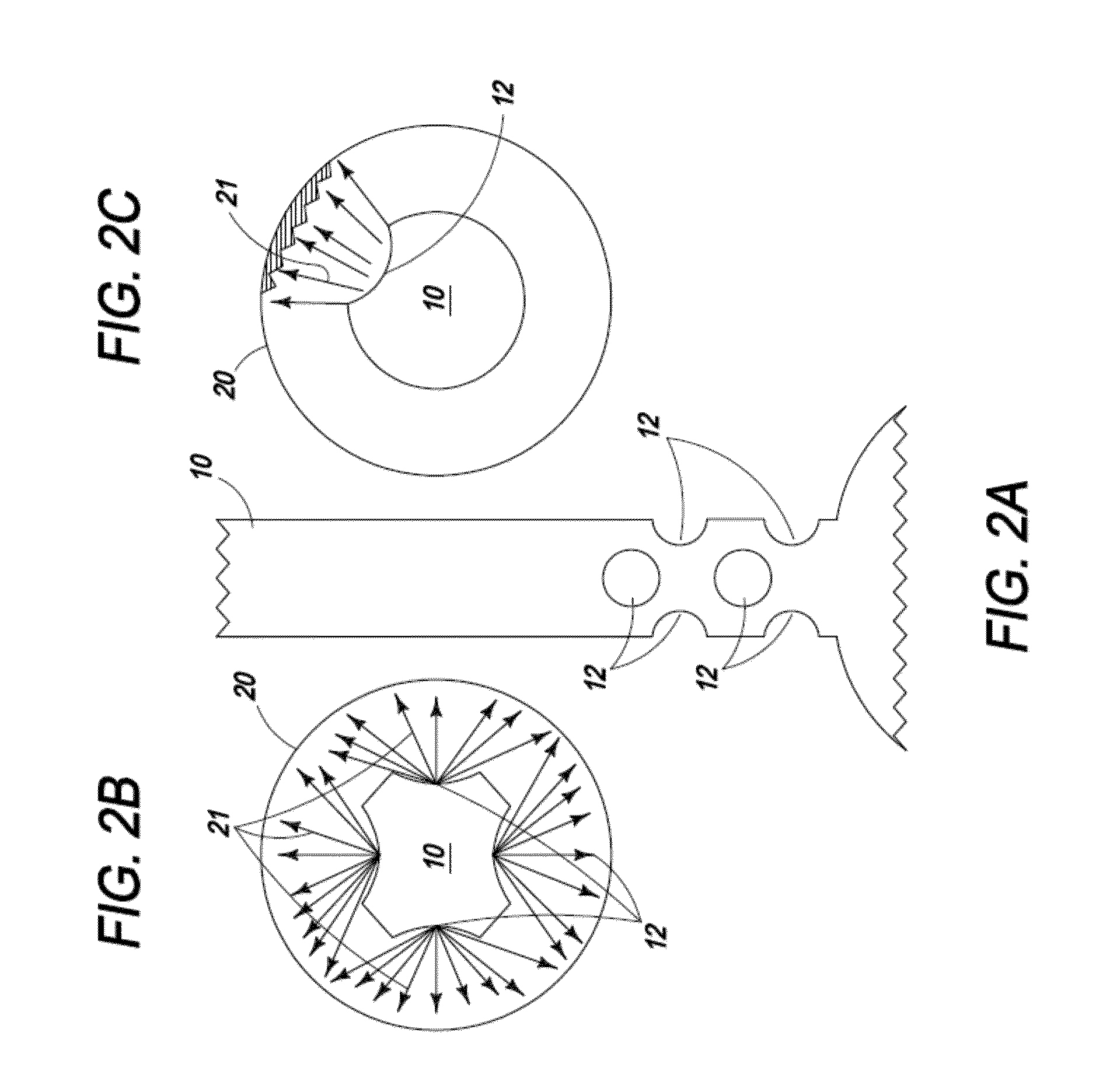
[0085]The present invention is directed toward an ablation device comprising a catheter with one or more centering or positioning attachments at one or more ends of the catheter to affix the catheter and its infusion port at a fixed distance from the ablative tissue which is not affected by the movements of the organ. The arrangement of one or more spray ports allows for uniform spray of the ablative agent producing a uniform ablation of a large area, such as encountered in Barrett's esophagus. The flow of ablative agent is controlled by the microprocessor and depends upon one or more of the length or area of tissue to be ablated, type and depth of tissue to be ablated, and distance of the infusion port from or in the tissue to be ablated.

[0086]The present invention is also directed toward a device to be used in conjunction with a tissue ablation system, comprising: a handle with a pressure-resistant port on its distal end, a flow channel through which an ablative agent can travel, ...


PUM


Abstract

The present application discloses devices that ablate human tissue. The device comprises a catheter with a shaft through which an ablative agent can travel, a liquid reservoir and a heating component, which may comprise a length of coiled tubing contained within a heating element, wherein activation of said heating element causes said coiled tubing to increase from a first temperature to a second temperature and wherein the increase causes a conversion of liquid within the coiled tubing to vapor, a reusable cord connecting the outlet of the reservoir to the inlet of the heating component, and a single use cord connecting a pressure-resistant inlet port of a vapor based ablation device to the outlet of the heating component.

Description

CROSS-REFERENCE TO RELATED APPLICATIONS[0001]This present application is a continuation in-part of U.S. patent application Ser. No. 12 / 573,939, filed on Oct. 6, 2009, entitled “Method and Apparatus for Tissue Ablation”, which relies on U.S. Provisional Patent Application No. 61 / 102,885, filed on Oct. 6, 2008, for priority, both of which are herein incorporated by reference.[0002]The present application also relies on U.S. Provisional Patent Application No. 61 / 493,344, entitled “Method and Apparatus for Tissue Ablation”, filed on Jun. 3, 2011, and assigned to the applicant of the present invention, for priority, which is herein incorporated by reference in its entirety.FIELD OF THE INVENTION[0003]The present invention relates to medical apparatus and procedures. More particularly, the present invention relates to a device for ablation of tissue comprising a centering or positioning attachment in order to position the device at a consistent distance from the tissue to be ablated.BACKG...

Claims


Application Information

Patent Timeline
IPC IPC(8): A61B18/04
CPCA61B5/1076A61B2017/4216A61B18/04A61B2017/00084A61B2018/048A61M25/1011A61M2205/3368A61B2018/00577A61B2018/00642A61B2018/00744A61B2018/00791A61B2019/464A61B5/6853A61B2017/00809A61B17/24A61B18/12A61B18/14A61B2018/0022A61B2018/00488A61B2018/00494A61B2018/00559A61B2090/064A61B2017/00203A61B2017/00973A61B2018/00327A61B2018/00541A61B2018/00547A61B2018/00648A61B2018/00714A61M25/04
Inventor SHARMA, VIRENDER K.PARKS, WILLIAM
Owner SANTA ANNA TECH LLC
Why Eureka
Try Eureka