A Fracture Healing Simulation Method Based on Cell Activity
A fracture healing and simulation method technology, applied in the field of biomedical engineering, can solve the problems that mechanical factors do not have a deterministic relationship between cell differentiation, cell proliferation, differentiation and apoptosis are not considered, and the setting of fracture healing areas is too simplified, so as to avoid Humanitarian controversies, effects on reducing delayed union and improving fracture healing quality
- Summary
- Abstract
- Description
- Claims
- Application Information
AI Technical Summary
Problems solved by technology
Method used
Image
Examples
specific Embodiment approach 1
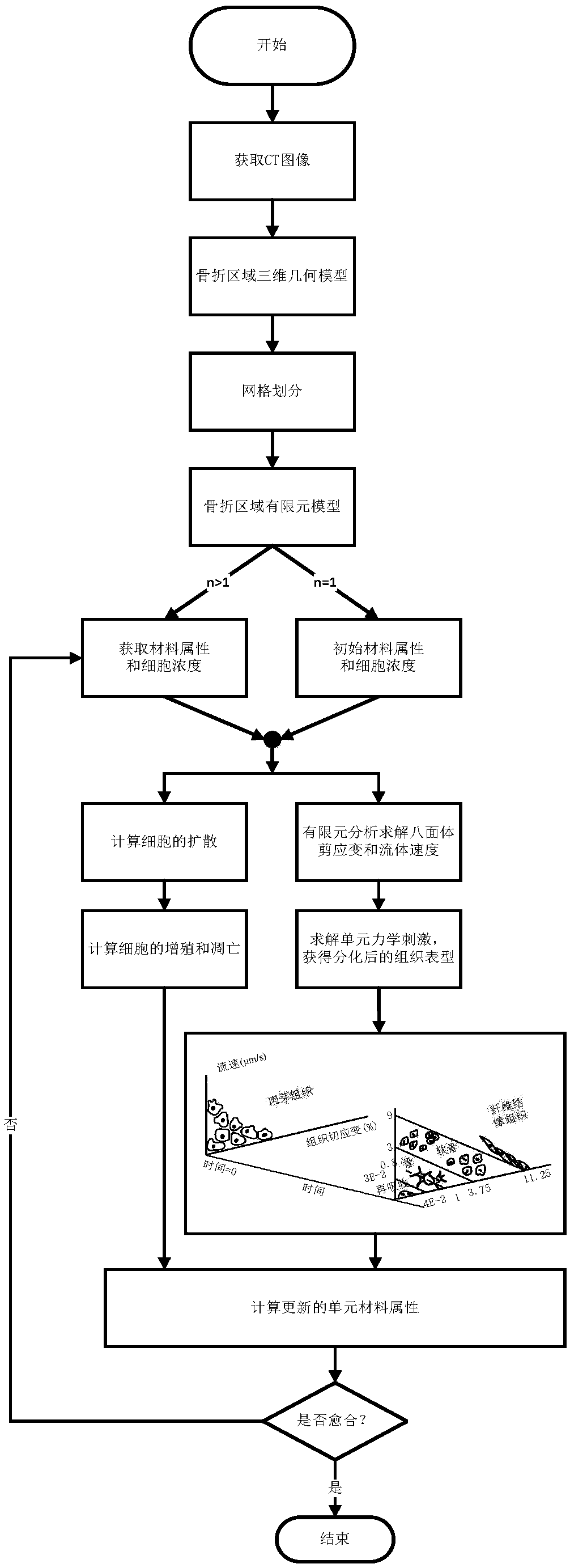
[0026] Specific embodiment 1: a kind of fracture healing simulation method based on cell activity comprises the following steps:
[0027] Step 1: Establish a three-dimensional geometric model of the fracture area;
[0028] Step 2: Mesh the 3D geometric model established in Step 1 to obtain a finite element model of the fracture area;
[0029] Step 3: Obtain the material properties and cell concentration of the fracture area unit;
[0030] Step 4: Finite element analysis of the fracture area, solving the octahedral shear strain and flow velocity suffered by the fracture area units;
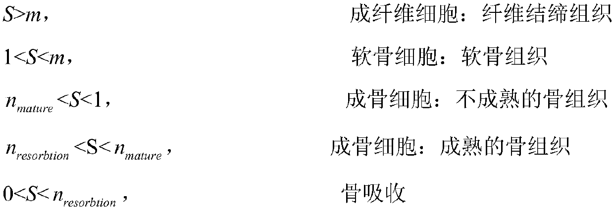
[0031] Step 5: Solve the mechanical stimulation of the fracture area unit, and obtain a new tissue phenotype according to the force adjustment algorithm;
[0032] Step 6: Analyze cell activity;
[0033] Step 7: Calculate the material properties of the new fracture area elements;
[0034] Step 8: Determine whether the program meets the termination condition. If not, the program enters the next i...
specific Embodiment approach 2
[0036] Specific embodiment two: the difference between this embodiment and specific embodiment one is: the establishment of the three-dimensional geometric model of the fracture area in the step one is specifically:
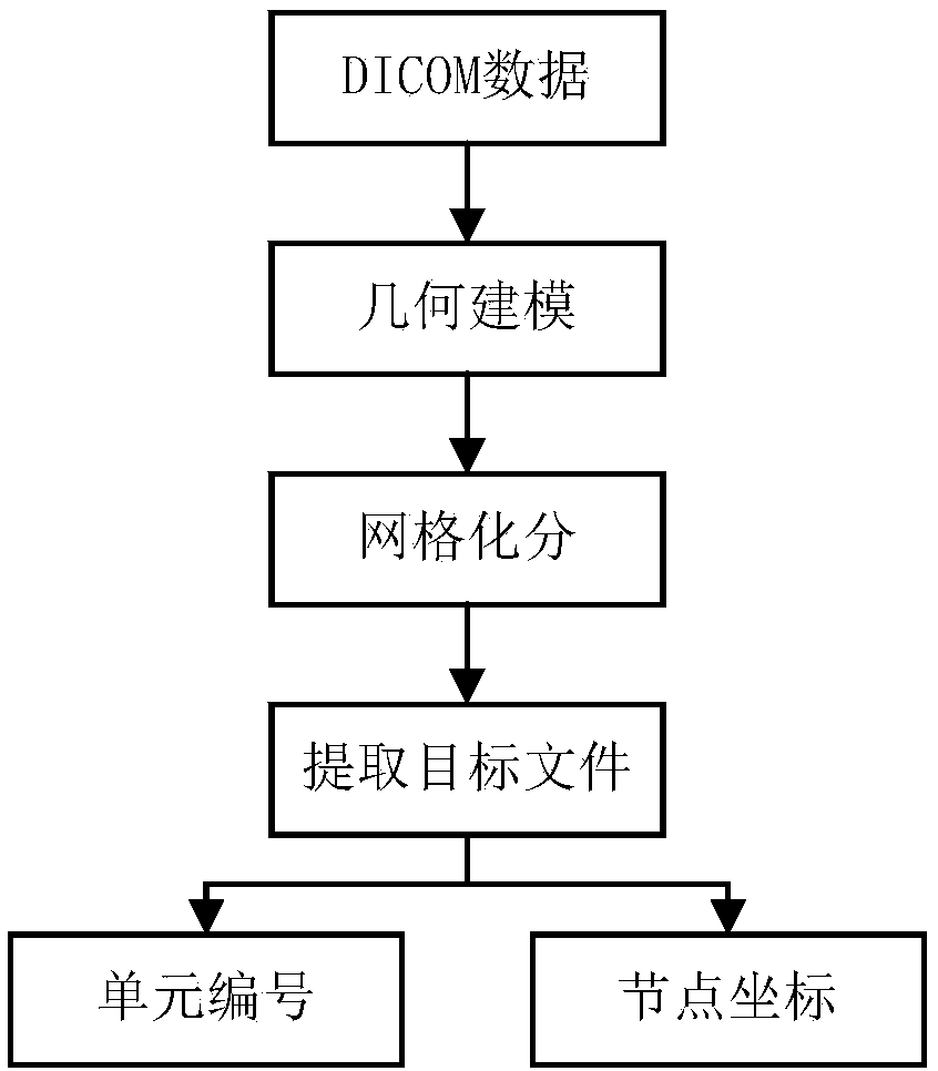
[0037] Using the segmentation-based 3D medical image surface reconstruction algorithm to reconstruct the 3D surface of the image to obtain a 3D geometric model;
[0038] The image is obtained by imaging equipment CT, and the data storage format is DICOM;
[0039] The process of constructing the solid model from the surface model is to construct the surface list of the solid model from the triangular slice sequence of the surface model and the upper and lower bottom surfaces, and construct the vertex list of the solid model from the vertex sequence of the surface model, and simultaneously establish the body, ring, edge and half edge The linked list and the pointing relationship of the nodes in each linked list. The solid construction process expressed by the boun...
specific Embodiment approach 3
[0041] Specific embodiment three: the difference between this embodiment and specific embodiment one or two is: the step two adopts octahedral mesh division unit to divide the three-dimensional geometric model into a grid, and the specific process of obtaining the finite element model of the fracture area is as follows:
[0042] Import the three-dimensional geometric model established in the specific embodiment one into the mesh division software, perform mesh division, and obtain the finite element model of the fracture area;
[0043] After meshing by meshing software, a lot of data will be obtained. Since only node coordinates and element numbers are needed to represent the finite element model of the fracture healing area, it is necessary to import the obtained data into MATLAB for preprocessing, and only extract the target data. According to the target data, two files of element number and node coordinates required for subsequent finite element calculation are generated;
...
PUM
 Abstract
Description
Claims
Application Information
 - R&D
- Intellectual Property
- Life Sciences
- Materials
- Tech Scout
- Unparalleled Data Quality
- Higher Quality Content
- 60% Fewer Hallucinations
Browse by: Latest US Patents, China's latest patents, Technical Efficacy Thesaurus, Application Domain, Technology Topic, Popular Technical Reports.
© 2025 PatSnap. All rights reserved.Legal|Privacy policy|Modern Slavery Act Transparency Statement|Sitemap|About US| Contact US: help@patsnap.com


