Blood analysis method and system

A blood flow analysis and technology to be analyzed, applied in the field of medical images, can solve the problems of unfamiliar operational engineering models and poor user experience

Inactive Publication Date: 2017-05-31
SHANGHAI UNITED IMAGING HEALTHCARE
View PDF6 Cites 21 Cited by
  • Summary
  • Abstract
  • Description
  • Claims
  • Application Information

AI Technical Summary

Problems solved by technology

[0004] Embodiments of the present invention provide a blood flow analysis method and system to achieve direct assessment of vascular parameters, thereby assisting doctors to accurately analyze patients' vascular diseases, and at the same ti

Method used


Image

  • Blood analysis method and system
  • Blood analysis method and system
  • Blood analysis method and system

Examples


Embodiment 1

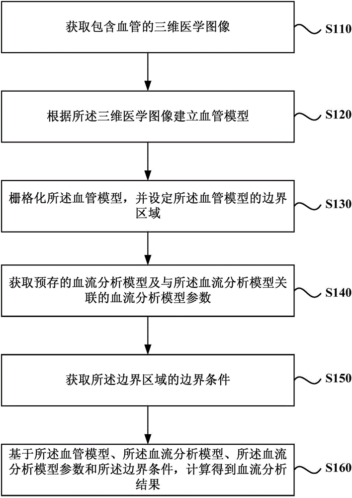
[0063] figure 1 It is a flowchart of a blood flow analysis method provided in Embodiment 1 of the present invention. This embodiment is applicable to the situation of analyzing and evaluating the parameters related to hemodynamics of blood vessels. The method can be executed by a blood flow analysis device or a blood flow analysis system, and the device or system can be realized by software and / or hardware. Typically, the device or system can be configured in CT equipment or magnetic resonance equipment. see figure 1 , the blood flow analysis method provided in this embodiment includes:

[0064] S110. Acquire a three-dimensional medical image including blood vessels.

[0065] Wherein, the blood vessel may be any blood vessel that is prone to vascular diseases, specifically, the blood vessel may be a cardiovascular or other blood vessel, such as a carotid artery, a cranial artery, a chest artery, an abdominal artery, or a brachial artery. or leg arteries etc. Medical imag...

Embodiment 2

[0109] Figure 5 It is a flowchart of a blood flow analysis method provided by Embodiment 2 of the present invention. This embodiment is an optional solution proposed on the basis of the foregoing implementation one. see Figure 5 , the blood flow analysis method provided in this embodiment includes:

[0110] S210. Acquire a three-dimensional medical image including blood vessels.

[0111] S220. If the blood vessel part is a common blood vessel part, use a preset automatic segmentation algorithm to automatically segment the blood vessel model corresponding to the blood vessel part from the three-dimensional medical image; otherwise, obtain the user's editing of the blood vessel part; according to the editing result A blood vessel model corresponding to the blood vessel part is segmented from the three-dimensional medical image.

[0112] Among them, common blood vessels can be coronary arteries, abdominal aneurysms, cerebral arteries, or lower extremity arteries.

[0113] ...

Embodiment 3

[0122] Figure 7 It is a flowchart of a blood flow analysis method provided in Embodiment 3 of the present invention. This embodiment is an optional solution proposed on the basis of the above embodiments, taking coronary vessels as an example. see Figure 7 , the blood flow analysis method provided in this embodiment includes:

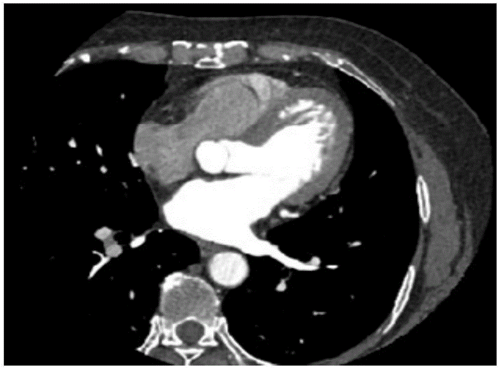
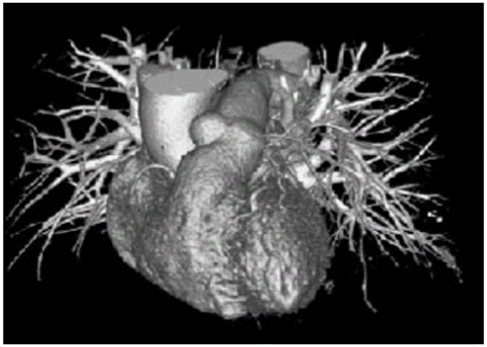
[0123] S310. Acquire a plurality of enhanced coronary CT images of the patient, and establish a three-dimensional medical image of the coronary artery according to the enhanced images.

[0124] Such as Figure 8a , 8b As shown in and 8c, the enhanced coronary CT images of the patient from different angles are collected, and a three-dimensional medical image of the coronary artery is established based on the enhanced images.

[0125] S320. Taking the entire coronary artery and a segment of the aorta as the blood vessel part to be analyzed, and extracting a blood vessel model corresponding to the blood vessel part from the three-dimensional medical...


PUM


Abstract

The embodiment of the invention discloses a blood analysis method and system, and relates to the technical field of medical hydromechanics and medical images. The method comprises the following steps of obtaining a three-dimensional medical image containing blood vessels; building a blood vessel model according to the three-dimensional medical image; performing rasterization on the blood vessel model; setting the boundary region of the blood vessel model; obtaining the prestored blood analysis model and a blood analysis model parameter relevant to the blood analysis model; obtaining the boundary condition of the boundary region; on the basis of the blood vessel model, the blood analysis model, the blood analysis model parameter and the boundary condition, performing calculation to obtain the blood analysis result. The blood analysis method and system provided by the embodiment of the invention have the advantages that the direct evaluation on the blood vessel parameter is realized; meanwhile, the problem of poor user experience due to unfamiliarity to the operation engineering model for direct evaluation on blood vessel parameters by users (doctors) is solved.

Description

technical field [0001] Embodiments of the present invention relate to the technical field of medical images, and in particular, to a blood flow analysis method and system. Background technique [0002] At present, there are more and more vascular diseases, such as loss of elasticity of blood vessel walls, stenosis of blood vessel lumen, and thrombus. In the prior art, vascular diseases can be monitored through non-invasive detection and invasive detection methods. Among them, non-invasive testing may include electrocardiography, blood test-based biomarker evaluation, tablet testing, echocardiography, computed tomography, and positron emission tomography; invasive testing may include diagnostic cardiac catheterization, among others. [0003] In the process of realizing the present invention, the inventors found that the defects of the prior art are: the above-mentioned test methods generally do not provide direct evaluation of the blood flow velocity, blood pressure, blood v...

Claims


Application Information

Patent Timeline
IPC IPC(8): G06T7/00G06T17/00G06T7/13
CPCG06T7/0012G06T17/00G06T2207/30104G06T2210/41
Inventor 马杰延任远
Owner SHANGHAI UNITED IMAGING HEALTHCARE
Why Eureka
Try Eureka