In-vitro detection method for quickly screening exon 5-8 mutation of P53 gene and application

A detection method, an in vitro detection technology, applied in the determination/inspection of microorganisms, biochemical equipment and methods, DNA/RNA fragments, etc., can solve the problem of not being able to determine the position of mutations in DNA fragments, and facilitate high-throughput processing , high detection sensitivity and low dosage

Inactive Publication Date: 2017-06-13
北京青航基因科技有限公司
View PDF3 Cites 2 Cited by
  • Summary
  • Abstract
  • Description
  • Claims
  • Application Information

AI Technical Summary

Problems solved by technology

The fly in the ointment is that when performing unknown mutation screening and scanning, only the presence or

Method used


Image

  • In-vitro detection method for quickly screening exon 5-8 mutation of P53 gene and application
  • In-vitro detection method for quickly screening exon 5-8 mutation of P53 gene and application
  • In-vitro detection method for quickly screening exon 5-8 mutation of P53 gene and application

Examples


Embodiment 1

[0031] An in vitro detection method for rapid screening of P53 gene exon 5-8 mutations, comprising the following steps:

[0032] (1) Extraction of Dried Blood Spot DNA

[0033] The total DNA extraction was carried out according to the instructions of the Magnetic Bead Method Dried Blood Spot Genomic DNA Extraction Kit (Tiangen), as follows:

[0034] 1. Take a new 96-well deep-well plate, and add three 3*3mm dried blood spot samples to each well. Add 200 µL of buffer GA to a 96 deep-well plate.

[0035] 2. Add 20 μL of proteinase K solution, vortex for 10 seconds to mix well, put it into a constant temperature oscillator preheated to 56 degrees, and lyse at 900 rpm for 30 minutes.

[0036] 3. During the sample lysis in the previous step, add 10 μL magnetic bead suspension G, 200 μL buffer GB and 200 μL absolute ethanol to a new 96-well deep-well plate, and mix by whipping or shaking for 10 seconds.

[0037]4. After the sample is lysed, briefly centrifuge the deep-well plate ...

Embodiment 2

[0056] Embodiment 2: clinical sample detection

[0057] The method of Example 1 was used to detect exons 5-8 of the P53 gene in the blood of 200 cases of physical examination population, and the mutation of the P53 gene in the blood DNA of 3 cases of subjects could be successfully detected, and then the PCR product was analyzed by using the next generation sequencing method. After detection and comparison with the human genome sequence, it was confirmed that there were indeed changes in the DNA sequence of these three samples.

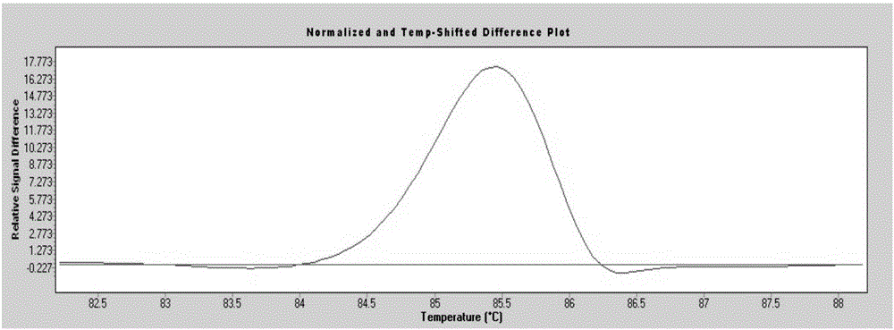
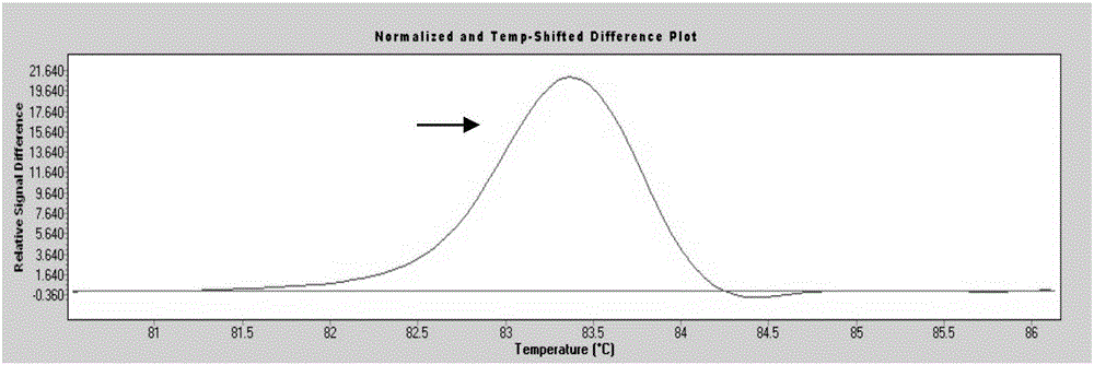
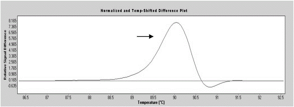
[0058] Among the 200 blood samples from the physical examination population, 3 cases (No. 68, No. 105, and No. 160) had mutations (separate peaks appeared), and No. 68 sample P53 gene exon 7 was suspected to have a nucleic acid sequence change ( figure 1 ); No. 105 sample P53 gene exon 6 is suspected to have a nucleic acid sequence change ( figure 2 ); Nucleic acid sequence change in exon 5 of P53 gene in sample No. 160 was suspected ( image 3 ). ...


PUM


Abstract

The invention provides an in-vitro detection method for quickly screening the exon 5-8 mutation of a P53 gene. The method comprises the following steps: (1) extracting the genome DNA of a peripheral blood sample; (2) designing and synthesizing 4 pairs of specific primers for amplifying exon 5-8 of the P53 gene; (3) performing high resolution melting curve analysis and grounding PCR on the genome DNA of the sample by using the primers; and (4) obtaining the sample with a positive result in the curve analysis, meaning occurrence of P53 gene mutation. The detection method is not limited by site or type of mutated base, does not need a sequence specific probe, and can directly run high resolution melting after PCR is completed to complete analysis on the sample mutation. The method is simple, convenient and quick in operation, is low in use cost and accurate in result, can realize truly closed pipe operation. Furthermore, the method has the characteristics of high through-put, high speed, kit availability, low detection cost and the like for quick screening of population.

Description

technical field [0001] The invention relates to a gene detection method. In particular, it relates to an in vitro detection method for rapid screening of P53 gene exon 5-8 mutation and its application. Background technique [0002] In recent years, the incidence and mortality of malignant tumors in my country have shown a clear upward trend, and have leapt to the top of the cause of death among Chinese residents. However, there is no breakthrough research on the early detection, early diagnosis and prevention of malignant tumors, and some patients are already in the middle and late stages when they see a doctor, and the curative effect is poor. Therefore, improving the early diagnosis level of malignant tumors is an effective way to improve the efficacy of malignant tumor diagnosis and treatment. At present, in clinical practice, serological detection methods are mostly used to monitor tumor markers in serum or other body fluids, and to dynamically make judgments on the ex...

Claims


Application Information

Patent Timeline
IPC IPC(8): C12Q1/68C12N15/11
CPCC12Q1/686C12Q1/6886C12Q2600/156
Inventor 曾瀚其他发明人请求不公开姓名
Owner 北京青航基因科技有限公司
Why Eureka
Try Eureka