Chest radiography anti-radiation X-ray camera system suitable for indoor radiology department

An X-ray and radiation protection technology, which is applied in the direction of radiological diagnosis equipment, radiation safety devices, applications, etc., can solve the problems of lack of effective radiation protection and affect the health of the examinee, so as to improve the overall protection quality and achieve better results. Radiation protection effect, easy-to-use effect

Pending Publication Date: 2017-06-23
南通市亿东医疗仪器有限公司
View PDF3 Cites 5 Cited by
  • Summary
  • Abstract
  • Description
  • Claims
  • Application Information

AI Technical Summary

Problems solved by technology

However, the current general-purpose X-ray imaging systems do not provide effective radiation protection for the non-examined position of the examinee, so that the examinee suffers from unexpected radiation damage and affects the health of the examinee.

Method used


Image

  • Chest radiography anti-radiation X-ray camera system suitable for indoor radiology department
  • Chest radiography anti-radiation X-ray camera system suitable for indoor radiology department

Examples


Embodiment Construction

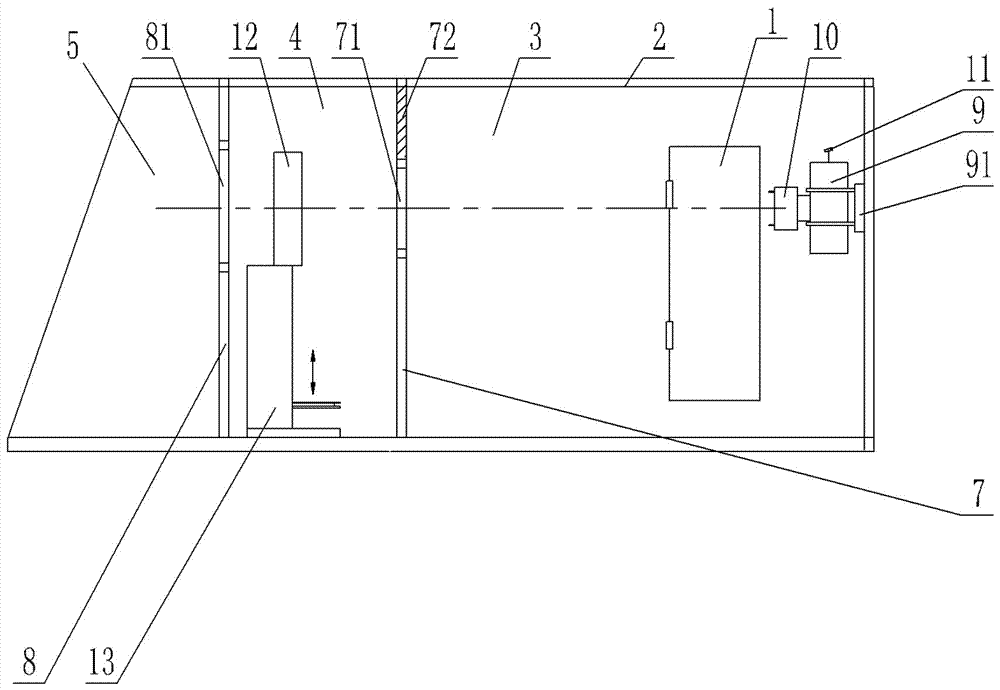
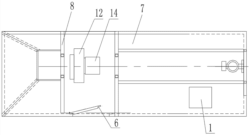
[0015] like figure 1 and figure 2 As shown, a chest radiation protection X-ray photography system suitable for indoor radiology, including a machine room 2, a front isolation board 7 and a rear isolation board 8 are arranged side by side in the machine room 2, and a 430×430mm opening is opened on the front isolation board 7 The front window 71 and the rear isolation plate 8 are provided with a 500×500mm rear window 81, and a physical examination area 4 is formed between the front isolation plate 7 and the rear isolation plate 8. The physical examination area 4 is provided with a radiation-proof door 6 for the physical examination area, and the physical examination area 4 There is a DR flat box 12 inside, and the lower edge of the rear window 81 should be slightly lower than the DR flat box 12 to ensure that the X-rays after photography can enter the X-ray leakage area 5 unobstructed, and the bottom of the DR flat box 12 is provided with a lifting box 13. After the subject st...


PUM


Abstract

The invention discloses a chest radiography anti-radiation X-ray camera system suitable for the indoor radiology department. The chest radiography anti-radiation X-ray camera system comprises a machine room, wherein a front isolation board and a rear isolation board are arranged in the machine room side by side, a front window is formed in the front isolation board, a rear window is formed in the rear isolation board, an emission area is formed in rear of the rear window, a physical examination area is formed between the front isolation board and the rear isolation board, an anti-radiation door of the physical examination area is arranged in the physical examination area, a DR flat-panel box is arranged in the physical examination area, a lifting box is arranged on the bottom of the DR flat-panel box, an X-ray area with the trapezoidal section is formed between the front isolation board and the top wall of the machine room, an X-ray bulb tube is arranged in the X-ray area, the X-ray bulb tube fixed to the rear wall of the machine room through a cushion plate, a light beam limiter is arranged at the emission end of the X-ray bulb tube, and the cross central lines of the light beam limiter, the front window, the DR flat-panel box and the rear window are overlapped. For the system provided by the invention, X-ray shooting is carried out on the position of an examined area by lifting an examined person, and the trapezoid-type lead plate protection of the X-ray area can effectively limit the emission direction of complement lines on the periphery of a main shooting beam of the X-rays; the X-rays penetrating through the rear window emit towards the tail and the bottom of the compartment after entering the emission area, and thus the integral protection quality is promoted.

Description

technical field [0001] The invention belongs to the field of X-ray photography radiation protection of medical equipment, and in particular relates to a chest X-ray radiation protection X-ray photography system suitable for indoor radiology departments. Background technique [0002] The currently known indoor X-ray photography system is composed of X-ray generator (high voltage generator), X-ray tube, light beam device, DR flat-panel photography device, display storage system, and control operating system. When in use, the subject stands in front of the DR panel, and the height of the X-ray tube is adjusted manually or electrically, and the position of the DR panel on its bracket is manually adjusted to adjust the ideal position of the subject. Using the characteristics of X-rays, the DR imaging flat panel receives signals and presents and retains the examinee's physical examination information through the display and storage system. However, none of the current general-pur...

Claims


Application Information

Patent Timeline
Patent Type & Authority Applications(China)
IPC IPC(8): A61B6/00A61B6/10E04H1/12
CPCA61B6/107A61B6/44E04H1/125
Inventor 蔡丽东蔡松汝
Owner 南通市亿东医疗仪器有限公司
Why Eureka
Try Eureka