Three-dimensional imaging method and system of organ and lesion based on medical images
A medical imaging and three-dimensional imaging technology, which is applied in the fields of instruments, image enhancement, image analysis, etc., to achieve the effect of convenient observation
- Summary
- Abstract
- Description
- Claims
- Application Information
AI Technical Summary
Problems solved by technology
Method used
Image
Examples
Embodiment 1
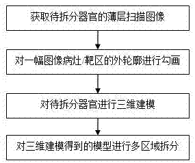
[0061] Embodiment 1 is the splitting of the brain lobe; in this embodiment, the organ to be split is the brain lobe, and the multiple regions are the frontal lobe, temporal lobe, parietal lobe, occipital lobe and cerebellum. The method includes the following sub-steps:
[0062] S11: Obtain a thin-layer scan image of the brain lobe of T1-weighted imaging;
[0063] T1-weighted imaging (T1-weighted imaging, T1WI) refers to this imaging method that focuses on the difference in longitudinal relaxation of tissues, while minimizing the influence of other tissue characteristics such as transverse relaxation on the image.
[0064] S12: outline the outer contour of the lesion / target area in one of the images;
[0065] S13: Carry out three-dimensional modeling of the lesion / target area and the organ to be split respectively, wherein for the three-dimensional modeling of the lesion / target area, the region growth algorithm with the same threshold is used to determine the boundary realizat...
Embodiment 2
[0072] Example 2 is the splitting of the liver. In this embodiment, the organ to be split is the liver, and the multiple regions are the left lobe and the right lobe of the liver; the method includes the following sub-steps:
[0073] S21: using DCMTK to read the DICOM sequence images of the liver;
[0074] Since the image storage and transmission of current medical imaging equipment is gradually moving closer to the DICOM standard, in the process of medical image processing, we often need to write various program modules related to DICOM format images to complete our own processing functions. It is a huge project to understand the DICOM protocol from scratch, and then write these codes to implement these protocols completely. The DCMTK developed by German offis company provides us with a platform to implement the DICOM protocol, so that we can easily complete our main work on the basis of it, without having to put too much energy on the details of the implementation of the DI...
Embodiment 3
[0090] Embodiment 3 is that the hospital has its own internal database and data processing center, specifically: the data center is set in the hospital and is connected to multiple doctor terminals in the hospital through the intranet. Each doctor's terminal connected to the thin-layer scanning instrument is connected to the data center inside the hospital through the intranet; the data center inside the hospital processes and saves the data inside the hospital. When the doctor needs a model, he can directly download send. Intranet connection is used to improve security performance.
PUM
 Abstract
Description
Claims
Application Information
 - R&D
- Intellectual Property
- Life Sciences
- Materials
- Tech Scout
- Unparalleled Data Quality
- Higher Quality Content
- 60% Fewer Hallucinations
Browse by: Latest US Patents, China's latest patents, Technical Efficacy Thesaurus, Application Domain, Technology Topic, Popular Technical Reports.
© 2025 PatSnap. All rights reserved.Legal|Privacy policy|Modern Slavery Act Transparency Statement|Sitemap|About US| Contact US: help@patsnap.com

