Transgenic lymphocyte co-expressing anti-EGFRvIII chimeric antigen receptor and nonfunctional EGFR receptor, and uses thereof
A chimeric antigen receptor, lymphocyte technology, applied in the field of biomedicine
- Summary
- Abstract
- Description
- Claims
- Application Information
AI Technical Summary
Problems solved by technology
Method used
Image
Examples
Embodiment 1
[0088] Cell lines and basic experimental techniques used in the embodiments of the present invention are as follows:
[0089] Generation of lentivirus and transduction of human T lymphocytes
[0090]Replication-defective lentiviral vectors were generated and collected by centrifugation for transduction of human T lymphocytes. The following is a brief introduction to the production and collection of lentiviral vectors: 293T cells were placed in a cell culture dish with a bottom area of 150-cm2, and Express-In (purchased from Open Biosystems / Thermo Scientific, Waltham , MA) for viral transduction of 293T cells. Add 15 μg of lentiviral transgenic plasmid, 5 μg of pVSV-G (VSV glycoprotein expression plasmid), 10 μg of pCMVR8.74 plasmid (Gag / Pol / Tat / Rev expression plasmid) and 174 μl of Express-In (concentration of 1 μg / μl). The supernatants were collected at 24 hours and 48 hours respectively, and centrifuged for 2 hours using an ultracentrifuge at 28,000 rpm (centrifuge roto...
Embodiment 2
[0095] Example 2 Construction of vectors co-expressing non-functional EGFR receptors and anti-EGFRvIII chimeric antigen receptors
[0096] In this example, the inventors cloned the sequence encoding the single-chain antibody against human EGFRvIII, the ζ-chain sequence of the 4-1BB intracellular segment and the T cell receptor combination into a lentiviral vector containing the EF-1 promoter ( lentiviral vector), during the cloning process, the selected restriction enzymes were XbaI and NotI double enzyme digestion, and NotI and XhoI double enzyme digestion. Lentiviral plasmid with antigen receptor complex (LV-EGFRvIIICAR). The sequence containing the synthetic IRES and expressing the non-functional EGFR receptor was cloned into the LV-EGFRvIII CAR vector plasmid to construct LV-EGFRvIII CAR / tEGFR. figure 1 is a schematic diagram of a lentiviral vector, including a sequence encoding an anti-EGFRvIII chimeric antigen receptor, an IRES, and a sequence encoding a non-functional ...
Embodiment 3
[0097] Example 3 Anti-EGFR Antibodies Can Effectively Kill and Eliminate T Lymphocytes Co-expressing Non-functional EGFR Receptor and Anti-EGFRvIII Chimeric Antigen Receptor
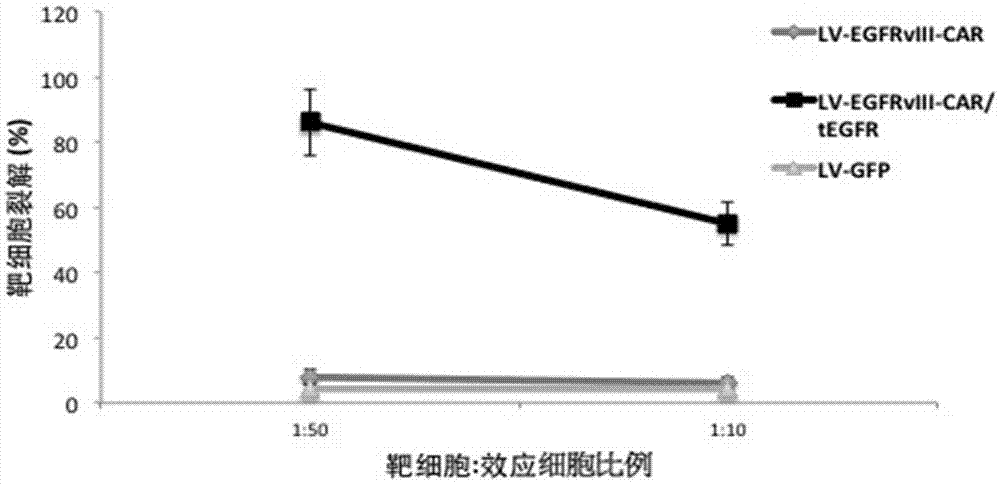
[0098] In this example, peripheral blood lymphocytes were obtained from anonymous blood donors. Peripheral blood lymphocytes were separated by gradient centrifugation using Ficoll-Hypaque. T lymphocytes and T cell activator magnetic beads CD3 / CD28 (purchased from Invitrogen, Carlsbad, CA) in 5% CO 2 , Incubated at 37 degrees Celsius for 72 hours, the medium was added with 2mmol / L glutamine, 10% high temperature inactivated fetal calf serum (FCS) (purchased from Sigma-Aldrich Co.) and 100U / ml of penicillin / chain RPMI medium 1640 (purchased from Invitrogen Gibco Cat. no. 12633-012) with antimycin double antibody. After activating and culturing for 72 hours, the cells were rinsed with washing solution to wash away the magnetic beads. The T cells were planted on cell culture dishes covered with recombinan...
PUM
 Abstract
Description
Claims
Application Information
 - R&D
- Intellectual Property
- Life Sciences
- Materials
- Tech Scout
- Unparalleled Data Quality
- Higher Quality Content
- 60% Fewer Hallucinations
Browse by: Latest US Patents, China's latest patents, Technical Efficacy Thesaurus, Application Domain, Technology Topic, Popular Technical Reports.
© 2025 PatSnap. All rights reserved.Legal|Privacy policy|Modern Slavery Act Transparency Statement|Sitemap|About US| Contact US: help@patsnap.com


