Hybridoma cell strain, anti-CRH antibody C-D17 based on hybridoma cell strain, and application of anti-CRH antibody C-D17 to detection

A hybridoma cell line and hybridoma cell technology are applied in the field of anti-adrenocorticotropin-releasing hormone monoclonal antibodies, which can solve problems such as health threats to rescue workers in plateau work, and achieve high sensitivity, low material consumption, and good specificity. Effect

Inactive Publication Date: 2017-08-18
陈锋 +1
1 Cites 0 Cited by

AI-Extracted Technical Summary

Problems solved by technology

If the prevention or treatment is not proper, acute mountain sickness will further develop into high altitude cerebral ...

Abstract

The invention relates to the field of biotechnology, and discloses an anti-CRH antibody C-D17 of a hybridoma cell strain, and application of the anti-CRH antibody C-D17 to detection. The hybridoma cell strain is preserved in China Center for Type Culture Collection on May 31, 2016 with the preservation number of CCTCC NO. C2015215, and is classified and named hybridoma cell C-D17. The monoclonal antibody C-D17 based on anti-cortin releasing hormone (CRH) generated by the hybridoma cell strain, and application of the monoclonal antibody to a kit for detecting acute altitude sickness are also provided. The anti-CRH monoclonal antibody C-D17 can recognize the C-end epitope of CRH, and has high specificity and high titer which can reach 1:4x10<6>. The double-antibody sandwich ELISA kit for detecting CRH has high specificity and sensitivity.

Application Domain

Technology Topic

HormoneDouble antibody sandwich +4

Image

  • Hybridoma cell strain, anti-CRH antibody C-D17 based on hybridoma cell strain, and application of anti-CRH antibody C-D17 to detection
  • Hybridoma cell strain, anti-CRH antibody C-D17 based on hybridoma cell strain, and application of anti-CRH antibody C-D17 to detection
  • Hybridoma cell strain, anti-CRH antibody C-D17 based on hybridoma cell strain, and application of anti-CRH antibody C-D17 to detection

Examples

  • Experimental program(4)

Example Embodiment

[0038] Example 1 Preparation and purification of the monoclonal antibody C-D17 of the present invention.
[0039] 1. Preparation of immunogen
[0040] 1.1 Preparation of CRHC synthetic peptide: Synthesize the C-terminal epitope of CRH, the sequence is the sequence shown in SEQ ID NO: 2, the purity is greater than 99%.
[0041] 1.2 Obtaining KLH-CRHC coupling peptide: Weigh 3 mg of meta-maleimide benzoic acid-N-hydroxysuccinimide ester (MBS) and dissolve in 200 μL dimethylformamide (DMF), take 1.5 mg KLH Dissolve in 0.5ml 0.05mol/L phosphate buffer (pH 7.0), add 70μL of the prepared MBS solution to it, and stir for 30min at room temperature; the KLH/MBS mixed solution is passed through the PD-10 desalting column, 0.05mol/L phosphate Buffer (pH 6.0) eluted and collected 3.5mL purified KLH/MBS; add 0.5mL deionized water, 5mg CRHC synthetic peptide dissolved in 100μL DMF, quickly add 1mL purified KLH/MBS, shake quickly and immediately use 2N Adjust the pH to 7.0-7.2 with NaOH, and place the mixed solution overnight at 4°C; finally add 3mL 0.1mol/LNH 4 HCO 3 , Freeze-drying to obtain KLH-CRHC coupling peptide, which is the immunogen used to prepare antibodies.
[0042] 2. Animal immunity and antiserum titer monitoring
[0043] Experimental animals: 6-week-old female Balb/c mice were purchased from Beijing Weitong Lihua Experimental Animal Company, the immune adjuvant Quick Antibody was purchased from Beijing Kangbiquan Biotechnology Co., Ltd., and mouse myeloma cells sp2/0 were stored by this unit.
[0044] 2.1 Immunization of mice: Take 50μg of KLH-CRHC conjugate peptide (in 250μL PBS), mix it with an equal volume of immune adjuvant by pipetting, and then 100μL each mouse, inject multiple points into the leg muscles; immunize once every 10 days .
[0045] 2.2 Titer detection: Take blood before immunization and on the 7th day after each immunization to monitor the titer of antiserum. The steps include:
[0046] (1) Coating: Dilute the KLH-CRHC coupling peptide (or KLH) to 10μg/mL with 0.05mol/L carbonate buffer (pH9.0) and add it to a 96-well polystyrene microplate. 100μL wells were coated overnight at 4°C, the liquid in the wells was discarded and washed 3 times with washing buffer PBST (0.05% Tween-20 in 0.02mol/L PBS), each time for 3 minutes;
[0047] (2) Blocking: add 300μL of BSA blocking solution to each well, block for 2h at 37°C, discard the liquid, and wash 3 times with washing buffer PBST (0.02mol/L PBS containing 0.05% Tween-20), each for 3 minutes;
[0048] (3) After diluting the serum sample of the test titer with BSA blocking solution, add 100μL per well to the above-mentioned enzyme-labeled plate, make three replicate wells for each dilution multiple, incubate at 37℃ for 1h, discard the liquid Wash with washing buffer PBST (0.05% Tween-20 in 0.02mol/LPBS) 3 times, 3 minutes each time;
[0049] (4) Dilute goat anti-mouse IgG/HRP 5000 times with enzyme-labeled antibody diluent (PBS+1%BSA), add 100μL per well to the above-mentioned enzyme-labeled plate, incubate at 37℃ for 0.5h, discard the liquid and use Washing buffer PBST (0.02mol/L PBS containing 0.05% Tween-20) wash 3 times, 3 minutes each time;
[0050] (5) Add 100μL of TMB substrate color developing solution to each well. After 10 minutes of color development at room temperature, add 50μL of stop solution (2mol/L H2SO4) to each well to stop the reaction, measure the 450nm absorbance value, and use the mouse serum before immunization as negative Control, take the measured value/control value ≥2.1 as positive to judge the immune serum titer;
[0051] At each blood sampling time point, each group of mice was subjected to intraorbital blood sampling, and the serum titer of the mice was detected by the above ELISA experiment. After four immunizations, the titer was the highest, and the titer was> 10 5 The immunized mice arrange cell fusion experiments.
[0052] 3. Cell fusion, cell line establishment and antibody preparation
[0053] 3.1 Experimental preparation: The day before fusion, one normal mouse was sacrificed, and mouse peritoneal fluid was removed to prepare mouse feeder cells; on the day of fusion, the titer reached (or exceeded) 10 5 After sacrifice, the spleen of the mouse was taken out under aseptic conditions, crushed and filtered through a sterile 200 mesh screen, placed in sterile saline by pipetting and washing, and then counted; myeloma cells SP2/0 cells Cultivate in RPMI-1640 medium, wash and count the same as spleen cells;
[0054] 3.2 Cell fusion: Mix the prepared SP2/0 and spleen cells thoroughly at a ratio of 1:10, and add 1 mL of the fusion agent (sterile PEG-4000, 50%) pre-preserved in a 37°C water bath within 1 min, and 37°C statically After 1 min, slowly add 5mL RPMI-1640 incomplete culture solution (pre-warmed at 37℃) along the tube wall within 2min, while gently rotating the centrifuge tube; then add preheated RPMI-1640 solution to 20mL to terminate the fusion, and centrifuge at 800rpm For 10 minutes, resuspend the pelleted cells in HAT medium and culture the cells in 96-well cell culture plates. Observe and record the cell growth status every day. On the 3rd day after the fusion, the HAT medium was replaced with half the medium, and the HT medium was replaced with half the medium on the 7th and 10th days, and then the complete medium (RPMI-1640 containing 15% fetal calf serum) was used for medium replacement.
[0055] 3.3 Establishment of positive hybridoma cell lines: Refer to "Principles and Techniques of In vitro Culture", select the culture supernatant with cell clusters in the culture plate for ELISA detection on 10-14 days after fusion, and coat KLH as a negative control. Take the cells in the positive cell wells, expand the culture to 48 wells, and then test the titer again. Select the cells that continue to be positive and use the limiting dilution subcloning method to culture them. Select the cells that are still positive after subcloning four times. The cell strain is stored in the China Common Microbial Species Collection and Management Center, and the preservation number is CCTCC NO.C2015215; the cells are cryopreserved and used to prepare ascites after expanded culture.
[0056] 3.4 Preparation of ascites: Take 8-week-old female Balb/c mice and inject 0.5 mL of liquid paraffin into the abdominal cavity. One week later, collect the expanded hybridoma cells. Each mouse will be injected with 2×10⁵ cells, starting from the 6th day. The mice began to produce ascites one after another. The mouse ascites was collected by centrifugation at 10,000 rpm for 15 min. The ascites was diluted with an equal volume of phosphate buffer (pH 7.4) and filtered through a 0.45 μm microporous membrane, and stored at 4°C for use or -20°C.
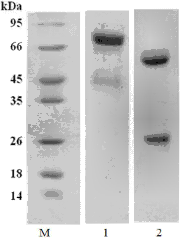
[0057] 3.5 Purification of monoclonal antibodies: Protein G affinity filler was purchased from Pharmacia, USA, and 10 mL of the stored ascites was purified with 2 mL of filler. The eluted antibody was desalted by Sephadex G-25, and the monoclonal antibody was determined by SDS-PAGE The purity of the antibody, C-D17 antibody purity reached more than 90%. The electrophoresis diagram of C-D17 after protein G affinity purification is shown figure 1.
[0058] 4. Characterization of monoclonal antibody C-D17
[0059] 4.1 Concentration determination: The concentration of the purified antibody was determined using Pierce's BSA protein quantification kit, C-D17 was 1.72 mg/mL.
[0060] 4.2 Subclass identification: The C-D17 monoclonal antibody purified from ascites is identified using the rapid antibody subclass identification test paper of Beijing Wukang Xinxing Technology Co., Ltd. The C-D17 antibody is of IgG1 type and the light chain is of κ type.
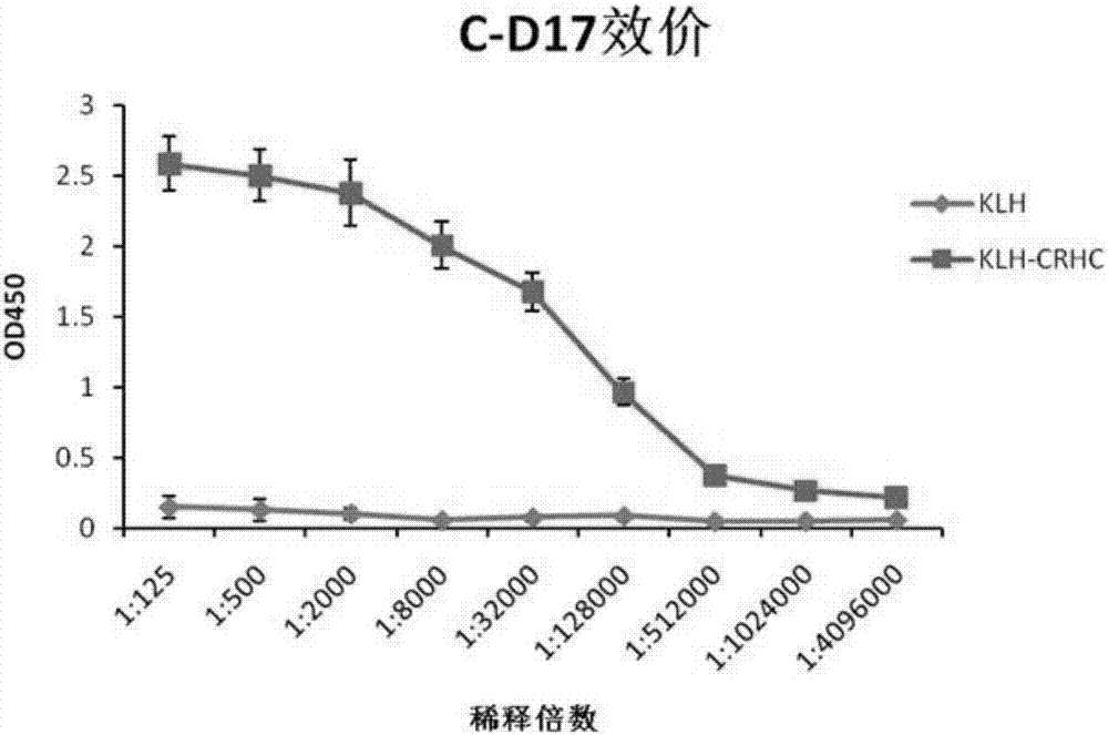
[0061] 4.3 Evaluation of titer: ELISA method is used to measure the titer of monoclonal antibody. Firstly, KLH-CRHC conjugated peptide and KLH were diluted to 10μg/mL with coating buffer respectively, and the ELISA plate was coated with 100μL/well. After coating at 37°C for 2h, after blocking with BSA blocking solution, add to each well Incubate the N-D19 antibody in multiple dilutions at 37°C for 1 hour, wash the plate, add 1:5000 diluted goat anti-mouse IgG/HRP, continue to incubate for 30 minutes, wash the plate thoroughly, add TMB color reagent to avoid light for 10 minutes, use 2mol/L H2SO4 terminates the color reaction, and read the absorbance at 450nm. See the result figure 2. figure 2 The results show that the titer of C-D179 antibody can reach 1:4×10 6.
[0062] 4.4 Identification of specificity and cross-reactivity: The indirect ELISA method is used to detect the specificity of C-D17 antibody and the cross-reactivity with KLH, KLH coupling peptide, CRH, luteinizing hormone (LH), etc., without immunized mouse serum Purified antibody is used as a control (Con), see the results of ELISA image 3. The results showed that C-D17 did not bind to any other proteins except KLH-CRHC and CRH, and the binding ability of C-D17 to KLH-CRHC was not significantly different from that of CRH.

Example Embodiment

[0063] Example 2 Preparation and purification of the monoclonal antibody N-D19 of the present invention.
[0064] 1. Preparation of immunogen
[0065] 1.1 Preparation of CRHN synthetic peptide: Synthesize the N-terminal epitope of CRH, the sequence is the sequence shown in SEQ ID NO:1, and the purity is greater than 99%.
[0066] 1.2 Obtaining KLH-CRHN coupling peptide: Weigh 3mg of meta-maleimidobenzoic acid-N-hydroxysuccinimide ester (MBS) and dissolve in 200μL dimethylformamide (DMF), take 1.5mg KLH Dissolve in 0.5ml 0.05mol/L phosphate buffer (pH 7.0), add 70μL of the prepared MBS solution to it, and stir for 30min at room temperature; the KLH/MBS mixed solution is passed through the PD-10 desalting column, 0.05mol/L phosphate Buffer (pH 6.0) eluted and collected 3.5mL purified KLH/MBS; add 0.5mL deionized water, 5mg CRHN synthetic peptide dissolved in 100μL DMF, quickly add 1mL purified KLH/MBS, shake quickly and immediately use 2N Adjust the pH to 7.0-7.2 with NaOH, and place the mixed solution overnight at 4°C; finally add 3mL 0.1mol/LNH 4 HCO 3 , Freeze-dried to obtain KLH-CRHN coupling peptide, which is the immunogen used to prepare antibodies.
[0067] 2. Animal immunity and antiserum titer monitoring
[0068] Experimental animals: 6-week-old female Balb/c mice were purchased from Beijing Weitong Lihua Experimental Animal Company, the immune adjuvant Quick Antibody was purchased from Beijing Kangbiquan Biotechnology Co., Ltd., and mouse myeloma cells sp2/0 were stored by this unit.
[0069] 2.1 Immunization of mice: Take 50μg of KLH-CRHN coupling peptide (in 250μL PBS), mix with an equal volume of immune adjuvant by pipetting, and then 100μL of each mouse, inject more than one leg muscle; immunize every 10 days .
[0070] 2.2 Titer detection: Take blood before immunization and on the 7th day after each immunization to monitor the titer of antiserum. The steps include:
[0071] (1) Coating: Dilute the KLH-CRHN coupling peptide (or KLH) to 10μg/mL with 0.05mol/L carbonate buffer (pH9.0) and add it to a 96-well polystyrene microplate. 100μL wells were coated overnight at 4°C, the liquid in the wells was discarded and washed 3 times with washing buffer PBST (0.05% Tween-20 in 0.02mol/L PBS), each time for 3 minutes;
[0072] (2) Blocking: add 300μL of BSA blocking solution to each well, block for 2h at 37°C, discard the liquid, and wash 3 times with washing buffer PBST (0.02mol/L PBS containing 0.05% Tween-20), each for 3 minutes;
[0073] (3) After diluting the serum sample of the test titer with BSA blocking solution, add 100μL per well to the above-mentioned enzyme-labeled plate, make three replicate wells for each dilution multiple, incubate at 37℃ for 1h, discard the liquid Wash with washing buffer PBST (0.05% Tween-20 in 0.02mol/LPBS) 3 times, 3 minutes each time;
[0074] (4) Dilute goat anti-mouse IgG/HRP 5000 times with enzyme-labeled antibody diluent (PBS+1%BSA), add 100μL per well to the above-mentioned enzyme-labeled plate, incubate at 37℃ for 0.5h, discard the liquid and use Washing buffer PBST (0.02mol/L PBS containing 0.05% Tween-20) wash 3 times, 3 minutes each time;
[0075] (5) Add 100μL of TMB substrate color developing solution to each well. After 10 minutes of color development at room temperature, add 50μL of stop solution (2mol/L H2SO4) to each well to stop the reaction, measure the 450nm absorbance value, and use the mouse serum before immunization as negative Control, take the measured value/control value ≥2.1 as positive to judge the immune serum titer;
[0076] At each blood sampling time point, each group of mice was subjected to intraorbital blood sampling, and the serum titer of the mice was detected by the above ELISA experiment. After four immunizations, the titer was the highest, and the titer was> 105 immunized mice arranged cell fusion experiments.
[0077] 3. Cell fusion, cell line establishment and antibody preparation
[0078] 3.1 Experimental preparation: The day before fusion, one normal mouse was sacrificed, and mouse peritoneal fluid was removed to prepare mouse feeder cells; on the day of fusion, the titer reached (or exceeded) 10 5 After sacrifice, the spleen of the mouse was taken out under aseptic conditions, crushed and filtered through a sterile 200 mesh screen, placed in sterile saline by pipetting and washing, and then counted; myeloma cells SP2/0 cells Cultivate in RPMI-1640 medium, wash and count the same as spleen cells;
[0079] 3.2 Cell fusion: Mix the prepared SP2/0 and spleen cells thoroughly at a ratio of 1:10, and add 1 mL of the fusion agent (sterile PEG-4000, 50%) pre-preserved in a 37°C water bath within 1 min, and 37°C statically After 1 min, slowly add 5mL RPMI-1640 incomplete culture solution (pre-warmed at 37℃) along the tube wall within 2min, while gently rotating the centrifuge tube; then add preheated RPMI-1640 solution to 20mL to terminate the fusion, and centrifuge at 800rpm For 10 minutes, resuspend the pelleted cells in HAT medium and culture the cells in 96-well cell culture plates. Observe and record the cell growth status every day. On the 3rd day after the fusion, the HAT medium was replaced with half the medium, and the HT medium was replaced with half the medium on the 7th and 10th days, and then the complete medium (RPMI-1640 containing 15% fetal calf serum) was used for medium replacement.
[0080] 3.3 Establishment of positive hybridoma cell lines: Refer to "Principles and Techniques of In vitro Culture", select the culture supernatant with cell clusters in the culture plate for ELISA detection on 10-14 days after fusion, and coat KLH as a negative control. Take the cells in the positive cell wells, expand the culture to 48 wells, and then test the titer again. Select the cells that continue to be positive and use the limiting dilution subcloning method to culture them. Select the cells that are still positive after subcloning four times. The cell line is stored in the China Common Microbial Species Collection and Management Center, and the preservation number is CCTCC NO.C2015216; the cells are cryopreserved and used to prepare ascites after expanded culture.
[0081] 3.4 Preparation of ascites: Take 8-week-old female Balb/c mice and inject 0.5 mL of liquid paraffin into the abdominal cavity. One week later, collect the expanded hybridoma cells. Each mouse will be injected with 2×10⁵ cells, starting from the 6th day. The mice began to produce ascites one after another. The mouse ascites was collected by centrifugation at 10,000 rpm for 15 min. The ascites was diluted with an equal volume of phosphate buffer (pH 7.4) and filtered through a 0.45 μm microporous membrane, and stored at 4°C for use or -20°C.
[0082] 3.5 Purification of monoclonal antibodies: Protein G affinity filler was purchased from Pharmacia, USA, and 10 mL of the stored ascites was purified with 2 mL of filler. The eluted antibody was desalted by Sephadex G-25, and the monoclonal antibody was determined by SDS-PAGE The purity of the antibody, N-D19 antibody purity reached more than 90%. The electropherogram of N-D19 after protein G affinity purification is shown Figure 4.
[0083] 4. Characterization of monoclonal antibody N-D19
[0084] 4.1 Concentration determination: The concentration of the purified antibody was determined using Pierce's BSA protein quantification kit, and the N-D19 was 2.33 mg/mL.
[0085] 4.2 Subclass identification: The N-D19 monoclonal antibody purified from ascites is identified using the rapid antibody subclass identification test paper of Beijing Wukang Xinxing Technology Co., Ltd. The N-D19 antibody is of IgG2a type and the light chain is of κ type.
[0086] 4.3 Evaluation of titer: ELISA method is used to measure the titer of monoclonal antibody. Firstly, KLH-CRHN coupling peptide and KLH were diluted to 10μg/mL with coating buffer respectively, and the microtiter plate was coated with 100μL/well. After coating at 37°C for 2h, after blocking with BSA blocking solution, add to each well Incubate the N-D19 antibody in multiple dilutions at 37°C for 1 hour, wash the plate, add 1:5000 diluted goat anti-mouse IgG/HRP, continue to incubate for 30 minutes, wash the plate thoroughly, add TMB color reagent to avoid light for 10 minutes, use 2mol/L H2SO4 terminates the color reaction, and read the absorbance at 450nm. See the result Figure 5. Figure 5 The results show that the titer of N-D19 antibody can reach 1:4×10 6.
[0087] 4.4 Identification of specificity and cross-reactivity: the indirect ELISA method is used to detect the specificity of N-D19 antibody and the cross-reactivity with KLH, KLH coupling peptide, CRH, luteinizing hormone (LH), etc., see the results of ELISA Image 6. The results showed that N-D19 did not bind to any other proteins except KLH-CRHN and CRH, and the binding ability of N-D19 to KLH-CRHN was not significantly different from that of CRH.

Example Embodiment

[0088] Example 3 The composition and optimal working concentration of the double antibody sandwich ELISA kit for detecting CRH of the present invention
[0089] 1. Preparation of HRP-labeled anti-human CRH monoclonal antibody C-D17 or N-D19
[0090] The method of HRP-labeled monoclonal antibody C-D17 or N-D19 is as follows: First, weigh 10 mg of dry HRP powder, dissolve it in 5 mL of double-distilled water, and add freshly prepared 0.2mol/L NaIO 4 0.5 mL of the solution, shake gently on a horizontal shaker at 4°C for 30 minutes, add 10 mg of the monoclonal antibody C-D17 or N-D19 antibody that has been dialyzed into carbonate buffer. After mixing, fill the solution with molecular weight cutoff In a 50kDa pre-treated dialysis bag, dialyze it against a 0.05mol/L carbonate buffer of pH 9.6 at 4°C, change the dialysate every 6h, and dialyze four times; after the dialysis, remove the dialysis Liquid in the bag, add NaBH with a concentration of 5mg/mL to it 4 100μL, 4°C, let stand for 4h; add an equal volume of saturated ammonium sulfate solution, 4°C overnight; centrifuge at 5000rpm for 10min, resuspend with 1mL 0.05mmol/L PBS (pH 7.4), and then add 0.05mmol/L PBS (pH 7.4) Buffer solution 4℃ for dialysis, change the dialysate every 6h, dialyze four times; take out the dialysis solution, add an equal volume of glycerol to the labeling solution to obtain purified HRP labeled monoclonal antibody C-D17 or N-D19.
[0091] 2. Label verification of enzyme-labeled antibody
[0092] ELISA method was used to detect the labeling effect of N-D19/HRP and C-D17/HRP. Coat the diluted C-D17 (or N-D19) (1μg/mL) on an enzyme-labeled plate (overnight at 4°C), block it with a blocking solution at 37°C for 2h, and add the diluted CRH antigen and N- D19/HRP (C-D17/HRP), continue to incubate for 1 hour, after washing the plate thoroughly, use TMB to develop color and stop the color reaction with the stop solution, and then read the OD450 (see Table 1). C-D17 antibody coating, N-D19 as the detection antibody is the best combination. In order to reduce the influence of background, the detection antibody concentration is 1:10000.
[0093] Table 1 CRH double antibody sandwich ELISA detection (OD450)
[0094]
[0095] 3. Detection limit and detection range of the kit
[0096] The detection sensitivity is 15.6pg/ml, and the best detection range is 15.6~1000pg/ml.
[0097] 4. Kit composition and detection method
[0098] The kit mainly includes: C-D19 pre-coated ELISA plate, PBST washing buffer, N-D19/HRP detection antibody, enzyme-labeled antibody diluent, TMB color developing solution, and stop solution.
[0099] The method of using the kit of the present invention includes the following steps:
[0100] (1) Dilute the serum collected by centrifugation twice, add it to the pre-coated ELISA plate, and place it at 37°C for 2 hours;
[0101] (2) Wash 3 times with PBST;
[0102] (3) Dilute the HRP-labeled antibody N-D19 10000 times with enzyme-labeled antibody diluent (PBS+1%BSA), add 100μL per well to the above-mentioned enzyme-labeled plate, incubate at 37℃ for 1h, discard the liquid and wash Wash with buffer PBST 3 times, 3 minutes each time;
[0103] (4) Add 100μL of TMB substrate color developing solution to each well. After 10 minutes of color development at room temperature, add 50μL of stop solution (2mol/L H2SO4) to each well to stop the reaction, and measure the 450nm absorbance.

PUM

no PUM

Description & Claims & Application Information

We can also present the details of the Description, Claims and Application information to help users get a comprehensive understanding of the technical details of the patent, such as background art, summary of invention, brief description of drawings, description of embodiments, and other original content. On the other hand, users can also determine the specific scope of protection of the technology through the list of claims; as well as understand the changes in the life cycle of the technology with the presentation of the patent timeline. Login to view more.

Similar technology patents

Classification and recommendation of technical efficacy words

  • High potency
  • Strong specificity

New Pyridine Analogues II

InactiveUS20070244088A1High potencyHigh selectivityAntibacterial agentsBiocidePyridineDisease injury
Owner:ASTRAZENECA AB

Cas9-foki fusion proteins and uses thereof

ActiveUS20150071899A1Strong specificityReduce the possibilityFusion with DNA-binding domainPeptide/protein ingredientsResearch settingNuclease
Owner:PRESIDENT & FELLOWS OF HARVARD COLLEGE

c-MET MODULATORS AND METHOD OF USE

InactiveUS20070179130A1Strong specificityEliminate side effectsBiocideSenses disorderSignal transductionCellular activity
Owner:EXELIXIS INC
Why Eureka
Try Eureka