
An immunofluorescent staining method and its kit for rapidly evaluating testicular spermatogenic function

A kit and reagent technology, applied in the field of reproductive medicine, can solve the problems of strong subjectivity and time-consuming, and achieve the effects of convenient operation, less time-consuming, and low detection cost

Active Publication Date: 2019-03-19
SHANGHAI FIRST PEOPLES HOSPITAL
View PDF5 Cites 0 Cited by
  • Summary
  • Abstract
  • Description
  • Claims
  • Application Information

AI Technical Summary

Problems solved by technology

[0004] The first purpose of the present invention is to provide a method for quickly and accurately evaluating the spermatogenic function of testicular tissue in view of the shortcomings of the prior art such as long time-consuming or strong subjectivity

Method used


Image

  • An immunofluorescent staining method and its kit for rapidly evaluating testicular spermatogenic function
  • An immunofluorescent staining method and its kit for rapidly evaluating testicular spermatogenic function
  • An immunofluorescent staining method and its kit for rapidly evaluating testicular spermatogenic function

Examples


Embodiment 1

[0037] Embodiment 1 detection kit

[0038] Reagent A: erythrocyte lysate;

[0039] Reagent B: 4% paraformaldehyde fixative;

[0040] Reagent C: PBS containing 5% fetal bovine serum (BSA) and 0.5% Triton X-100

[0041] Reagent D1: FITC-labeled rabbit anti-human GFRA1 antibody; PE-labeled mouse anti-human c-KIT antibody;

[0042] Reagent D2: FITC-labeled rabbit anti-human SCP3 antibody; PE-labeled mouse anti-human PNA antibody;

[0043] Reagent D3: 488 donkey anti-rabbit fluorescent secondary antibody; 555 donkey anti-mouse fluorescent secondary antibody;

[0044] Reagent E: fluorescent mounting medium;

[0045] Reagent F: PBS solution.

Embodiment 2

[0046] Embodiment 2 detection steps

[0047] Reagent preparation stage: Dilute reagents D1, D2, and D3 with reagent C at a certain ratio (recommended 1:500, which can be adjusted according to the staining situation) to make working solutions.

[0048] Stages of seminiferous tubule slide preparation:

[0049] (1) Obtain human seminiferous tubules (the length of which is 3-5cm can be sufficient for detection), put them into the upper filter of the improved EP tube, and rinse in reagent F (PBS solution) for 3 minutes;

[0050] (2) Take out the filter screen and discard the waste liquid in the lower layer, add 1ml of reagent B, and incubate at room temperature for 10 minutes;

[0051] (3) (Optional step, used when the obtained testicular tissue contains blood clots and a lot of blood) Take out the filter screen and discard the waste liquid in the lower layer, add 1ml of reagent A, and place on a shaker at room temperature for 10 minutes;

[0052] (4) Take out the filter screen a...

Embodiment 3

[0058] Embodiment 3 detection method

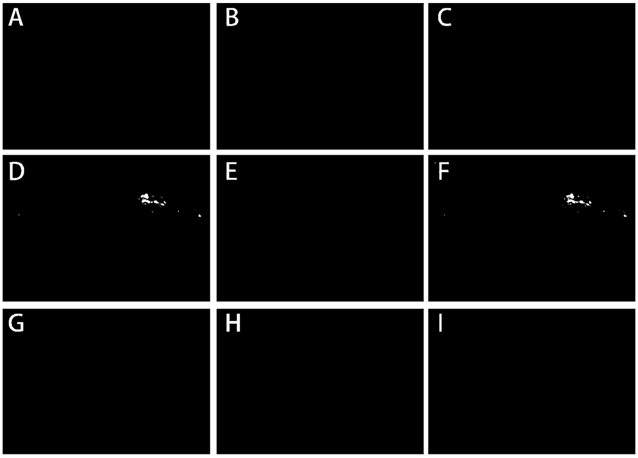
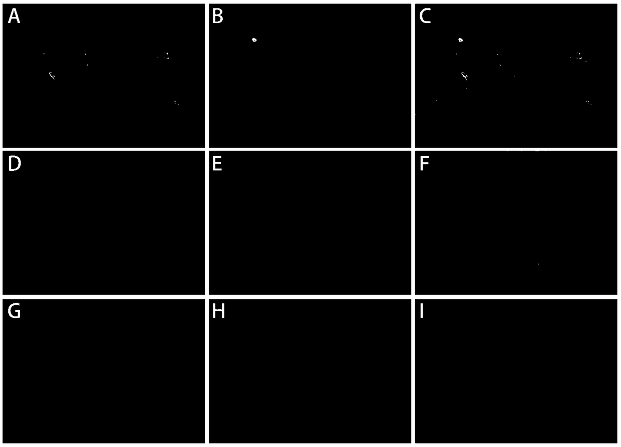
[0059] Use fluorescently labeled IgG antibodies to specifically bind to the currently recognized marker proteins of spermatogonial stem cells, differentiated spermatogonia, spermatocytes, and sperm (or sperm cells), and determine testicular spermatogenesis by observing the staining results The number and distribution of various spermatogenic cells in the tube can be used to determine the spermatogenic function of the patient's testis. In the seminiferous tubules incubated with reagent D1: the undifferentiated spermatogonia surface marker GFRA1 excited green fluorescence under the fluorescence microscope, and the differentiated spermatogonial cell surface marker c-KIT excited green fluorescence under the fluorescence microscope; the reagent D2 incubated In the seminiferous tubule: the pre-meiotic marker SCP3 of spermatocytes excites green fluorescence under the fluorescence microscope, and the sperm (sperm cell) acrosome marker PNA excites...


PUM


Abstract

The invention provides an antibody combination, a method and a detection kit for rapidly evaluating a testicular spermatogenic function. The antibody combination comprises (1) an FITC (fluorescein isothiocyanate) fluorescently-labeled rabbit antihuman GFRA1 antibody and a PE (phycoerythrin) fluorescently-labeled mouse antihuman c-KIT antibody, and (2) an FITC fluorescently-labeled rabbit antihuman SCP3 antibody and a PE fluorescently-labeled mouse antihuman PNA antibody. The method comprises the steps of taking a testiculus spermatogenic tubule as a detection sample, detecting expression conditions of GFRA1, c-KIT, SCP3 and PNA of the sample by immunohistochemical fluorescent staining, judging whether all spermatogenic cells exist, and then evaluating the spermatogenic function of a patient. According to the antibody combination, the method and the detection kit, immunofluorescent chemical staining of a germ cell surface marker is used for rapidly evaluating the spermatogenic function of the patient during / after an operation for the first time; the detection speed is high; the sensitivity is very high; the specificity is reliable; the spermatogenic cells in each stage can be distinguished; and a value for evaluation and subsequent treatment of the patient is important.

Description

technical field [0001] The invention relates to the technical field of reproductive medicine, in particular to an antibody combination, a staining method and a kit for rapidly and accurately evaluating the spermatogenic function of testicular tissue. Background technique [0002] The incidence of non-obstructive azoospermia (NOA) is about 10% in infertile men, accounting for 60% of azoospermia, and its characteristic is different degrees of spermatogenic damage in testicular tissue. The pathological classification of testicular tissue in patients with non-obstructive azoospermia is mainly divided into: 1. Low spermatogenicity; 2. Spermatogenic block; 3. Sertoli cell-only syndrome; etc. At present, the main method for judging the spermatogenesis of a patient during surgery (including testicular puncture, testicular biopsy, and testicular microsperm extraction) is to cut the testicular tissue into pieces as much as possible in HTF fluid (a human fallopian tube fluid analogue)....

Claims


Application Information

Patent Timeline
Patent Type & Authority Patents(China)
IPC IPC(8): G01N33/58G01N1/30G01N21/64
CPCG01N1/30G01N21/6428G01N33/582
Inventor 赵亮宇姚晨成李铮李朋孙红芳田汝辉万众陈慧兴杨超朱子珏
Owner SHANGHAI FIRST PEOPLES HOSPITAL
Learn More