Preparation containing fibroblast exosome and application thereof
A technology of fibroblasts and exosomes, which can be used in medical preparations containing active ingredients, aerosol delivery, skin diseases, etc., and can solve the problems of undisclosed fibroblast exosomes
- Summary
- Abstract
- Description
- Claims
- Application Information
AI Technical Summary
Problems solved by technology
Method used
Image
Examples
Embodiment 1
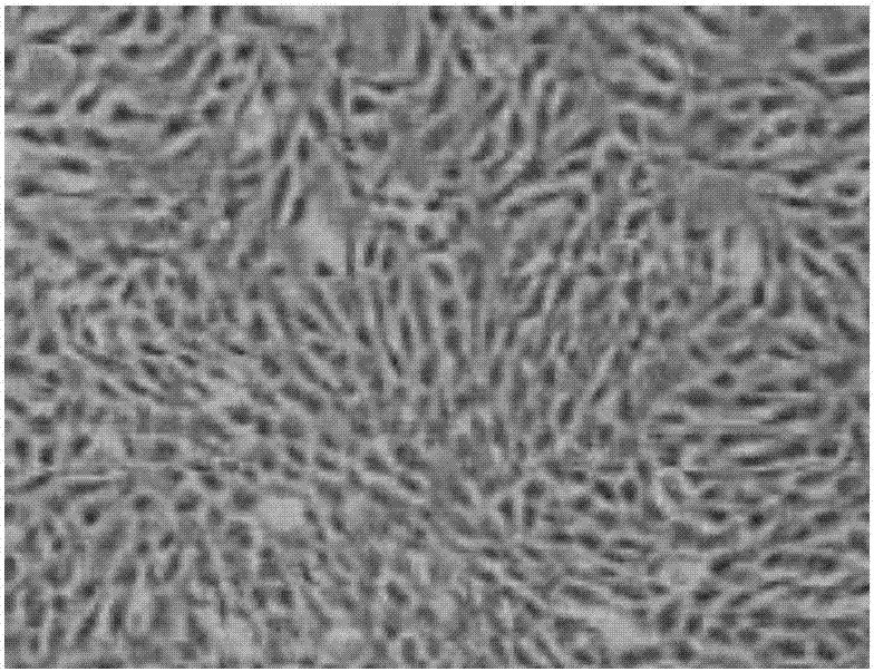
[0041] Take fresh healthy umbilical cord and umbilical cord blood, collect the umbilical cord blood and centrifuge, take the upper serum and inactivate it for later use. After rinsing with PBS, remove the umbilical vessels with scissors and tweezers, peel off the Wahrenheit jelly tissue inside, and cut the resulting tissue to a size of 1mm3, add α-MEM culture medium and place at 37°C, 5% CO 2 Culture in an incubator with 10% inactivated umbilical cord serum in the culture medium. After 5-8 days of umbilical cord tissue culture, it can be seen that some cells crawled out from around the tissue block, showing a small spindle shape. After one week, the cells began to proliferate rapidly and formed cell colonies of various sizes. After the cells were full, use 0.25 % Trypsinization for passage. Such as figure 1 As shown, the human umbilical cord mesenchymal stem cells grow in the form of fibroblast-like adherence, and the cells are uniform in shape, spindle-shaped, and have a ce...
Embodiment 2
[0043] Take the UC-MSCs isolated and cultured at passages P3 and P6 respectively, and adjust the cell density to 1x10 6 / ml, respectively incubated with FITC-CD29, PE-CD31, FITC-CD40, PE-CD44, PC5-CD45, PC5-CD90, PE-CD105 and FITC-HLA-DR antibodies at 4°C for 30 min, washed 3 times with PBS , and then resuspended the cells in 200 μl PBS, and detected on the flow cytometer.
[0044] As shown in Table 1, UC-MSCs highly express CD29, CD44, CD90, and CD105, and have low or no expression of CD31, CD40, CD45, and HLA-DR, which meet the identification criteria of UC-MSCs.
[0045] Table 1 Identification of UC-MSC surface markers
[0046]
[0047]
Embodiment 3
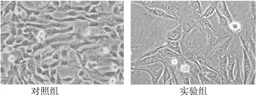
[0049] Take P6 generation UC-MSC, adjust the cell density to 1x10 6 / ml, according to 2x10 per bottle 6 Cells were seeded into T175 cell culture flasks. After 24 hours, the medium was changed and divided into two groups, namely the experimental group and the control group. The control group was added with normal medium (α-MEM containing 10% inactivated cord blood serum by volume fraction), and the experimental group was added with conditioned medium (α-MEM containing 10% volume fraction of inactivated cord blood serum, 10ng / ml basic fibroblast growth factor, 1mmol / L vitamin C phosphate, 0.04mmo / L proline) for cell induction, and morphological observation of the cells after 3 days .
[0050] Such as figure 2 As shown, the cells in the experimental group formed a flattened spindle-shaped or star-shaped structure, which conformed to the morphological characteristics of fibroblasts.
PUM
 Abstract
Description
Claims
Application Information
 - R&D
- Intellectual Property
- Life Sciences
- Materials
- Tech Scout
- Unparalleled Data Quality
- Higher Quality Content
- 60% Fewer Hallucinations
Browse by: Latest US Patents, China's latest patents, Technical Efficacy Thesaurus, Application Domain, Technology Topic, Popular Technical Reports.
© 2025 PatSnap. All rights reserved.Legal|Privacy policy|Modern Slavery Act Transparency Statement|Sitemap|About US| Contact US: help@patsnap.com


