
Medical instruments and techniques for treating pulmonary disorders

a technology of medical instruments and pulmonary disorders, applied in the field of tissue energy application methods and systems, can solve the problems of air-trapping and hyperinflation of the lungs, difficult breathing, and difficulty for the body to obtain oxygen and to get rid of carbon dioxide, so as to reduce the volume of the lung, enhance tissue remodeling, and cause the effect of lung volume reduction

Active Publication Date: 2011-02-22
TSUNAMI MEDTECH
View PDF234 Cites 110 Cited by
  • Summary
  • Abstract
  • Description
  • Claims
  • Application Information

AI Technical Summary

Benefits of technology

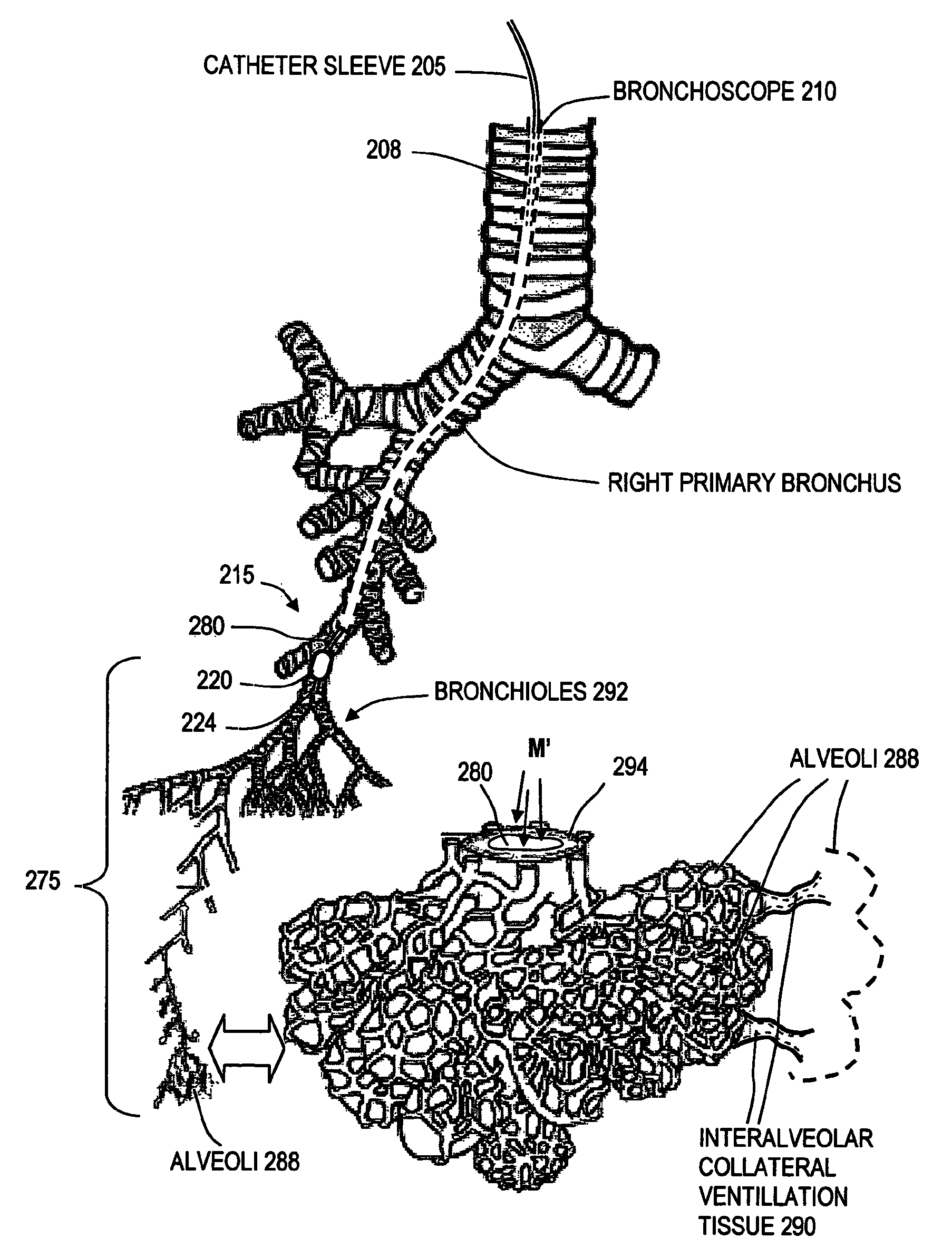
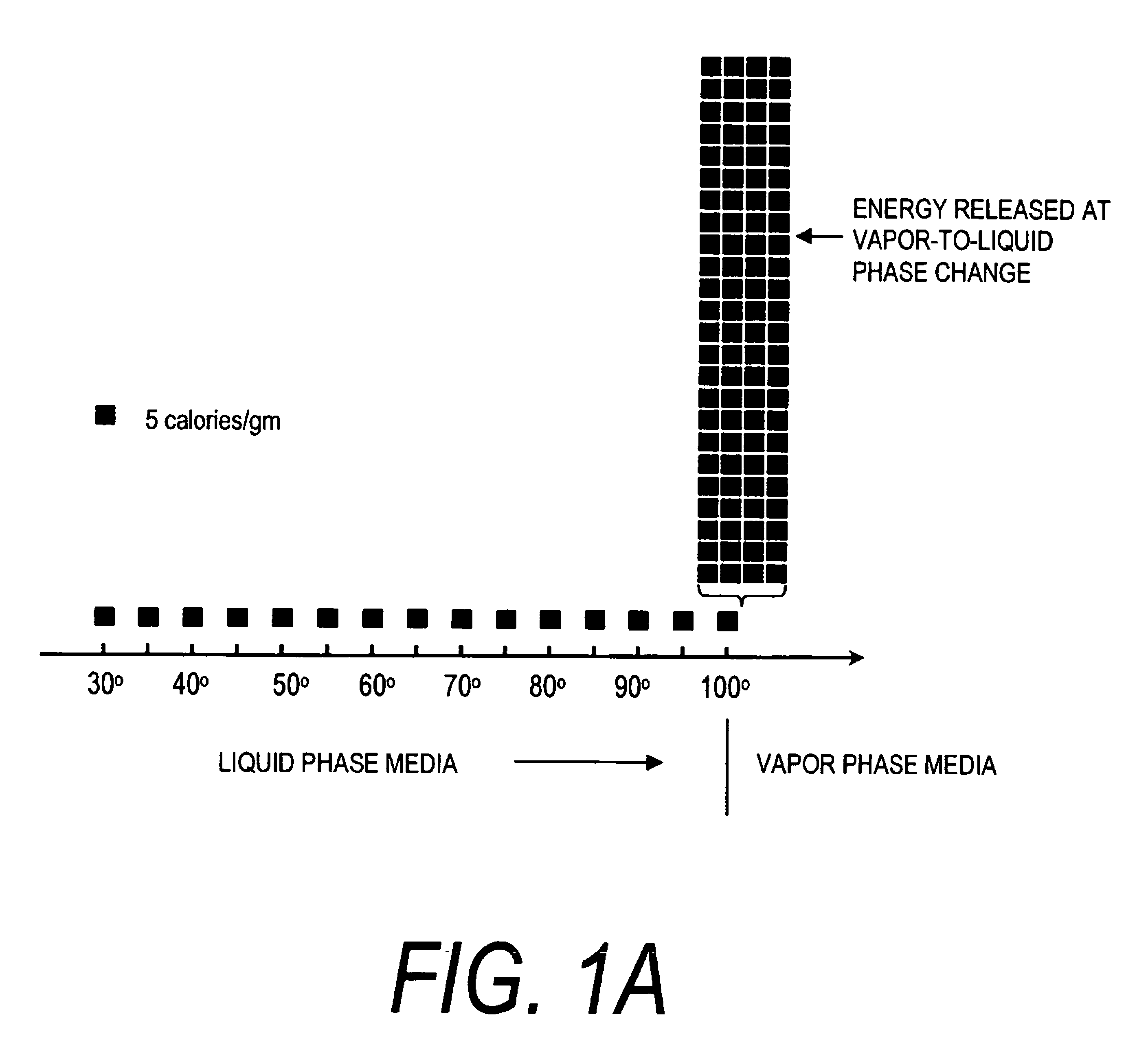
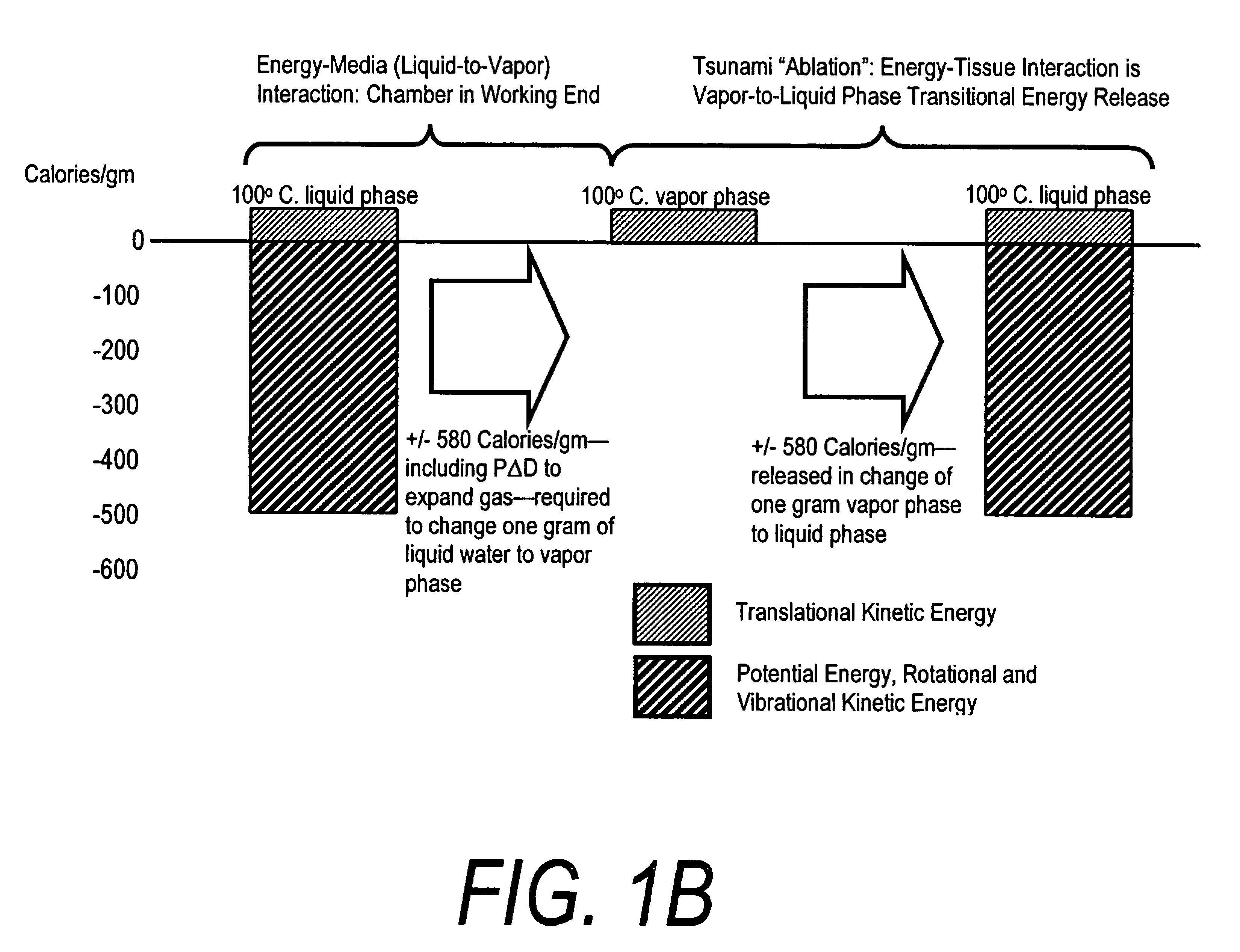
[0016]In general, the system of the invention is adapted to provide least invasive methods for lung volume reduction that are accomplished by using thermal energy to treat and shrink targeted regions of a the bronchial. In one embodiment, an elongated catheter is configured for introduction in a targeted airway. The handle portion of the catheter includes an interior chamber that is supplied with a biocompatible liquid under pressure. An energy source is coupled to the interior chamber to cause a liquid-to-vapor phase change in the biocompatible liquid, which contemporaneously ejects a flow of vapor from the working end of the catheter. The flow of vapor is controlled by a controller to cause a selected pressure and selected volume of vapor to propagate to alveoli. The vapor flow instantly undergoes a vapor-to-liquid phase change to thereby applying energy to the airway tissue. The thermal energy delivered is equivalent to the heat of vaporization of the fluid media, which shrinks and collapses the treated airways that are not supported by substantial cartilage. The treated tissue is maintained in a collapsed state by means of aspiration for a short interval to enhance tissue remodeling. Thereafter, the patient's wound healing response will cause fibrosis and further remodeling of the treated airway tissue to cause lung volume reduction.
[0017]The invention provides a method for LVR that can eliminate the complications of open surgery, endoscopic surgery or various bronchial plugs.
[0018]The invention provides a method for LVR that does not require transection of the exterior lung wall thus eliminating the serious complications of air leakage into the chest cavity.
[0019]The invention provides a method for LVR that can greatly reduce the patient's recuperative period and hospital stay.
[0021]The invention provides a method for LVR that will allow for greatly reduced costs when compared to open or endoscopic LVR procedures.

Problems solved by technology

Destruction of these air sacs makes it difficult for the body to obtain oxygen and to get rid of carbon dioxide.
The damage to the microscopic air sacs of the lung results in air-trapping and hyperinflation of the lungs.
As the damaged air sacs enlarge, they push on the diaphragm making it more difficult to breathe.
These changes produce the major symptom emphysema patients suffer—dyspnea (shortness of breath) and difficulty of expiration.
LVR was first introduced in the 1950's but was initially abandoned due to a high operative mortality, primarily due to air leakage.
One of the main difficulties of the procedure is suturing the resected lung margin in an airtight manner.
If an air leak allows air in the potential space between the ribs and lungs—then the vacuum effect will disappear and the lungs will sag upon chest expansion making it increasingly difficult to inflate the lungs and perform gas exchange.
As in any major thoracic procedure, there are many risks, including fever, wound infections, wound hematomas, postoperative fatigue and tachycardia.
Further, the duration of the improvement in lung function following resection is not yet completely known—but there is a suggestion that lung function begins to decline two years after LVR.
Despite optimistic reports, the morbidity, mortality and financial costs associated with LVR appear to be high, with some studies indicating mortality rates ranging from 4 to 17%.

Method used


Image

  • Medical instruments and techniques for treating pulmonary disorders
  • Medical instruments and techniques for treating pulmonary disorders
  • Medical instruments and techniques for treating pulmonary disorders

Examples


Embodiment Construction

[0038]1. Type “A” Thermotherapy Instrument. Referring to FIGS. 2A, 2B and 3, the working end 10 of a Type “A” system 5 of the present invention is shown that is adapted for endoscopic procedures in which a tissue volume T targeted for treatment (a thermoplasty) can be captured by a loop structure. The working end 10 comprises a body 11 of insulator material (see FIG. 3) coupled to the distal end of introducer member 12 extending along axis 15. In this exemplary embodiment, the working end 10 has a generally cylindrical cross-section and is made of any suitable material such as plastic, ceramic, glass, metal or a combination thereof. The working end 10 is substantially small in diameter (e.g., 2 mm to 5 mm) and in this embodiment is coupled to an elongate flexible introducer member 12 to cooperate with a working channel in an endoscope. Alternatively, the working end 10 may be coupled to a rigid shaft member having a suitable 1 mm to 5 mm or larger diameter to cooperate with a trocar...


PUM


Abstract

A surgical instrument for delivering energy to lung tissue, for example to cause lung volume reduction. In one embodiment, an elongated catheter has a handle portion that includes an interior chamber that is supplied with a biocompatible liquid media under pressure. An energy source delivers energy to the media to cause a liquid-to-vapor phase change within the interior chamber and ejects a flow of vapor media from the working end of the catheter. The delivery of energy and the flow of vapor are controlled by a computer controller to cause a selected pressure and selected volume of vapor to propagate to the extremities of the airways. Contemporaneously, the vapor undergoes a vapor-to-liquid phase transition which delivers a large amount of energy to airway tissue. The thermal energy delivered is equivalent to the heat of vaporization of the fluid media, which shrinks and collapses the treated airways. The treated tissue is the maintained in a collapsed state by means of aspiration for a short interval to enhance tissue remodeling. Thereafter, the patient's wound healing response causes fibrosis and further remodeling to cause permanent lung volume reduction.

Description

CROSS-REFERENCE TO RELATED APPLICATIONS[0001]This application claims benefit of U.S. Provisional Patent Application No. 60 / 615,900 filed Oct. 5, 2004 titled Medical Instruments and Techniques for Thermally-Mediated Procedures. This application is also a continuation-in-part of U.S. patent application Ser. No. 10 / 346,877 filed Jan. 18, 2003 now U.S. Pat. No. 6,911,028 titled Medical Instrument Working End and Method for Endoluminal Treatment, the entire contents of which is incorporated herein by this reference and should be considered a part of this specification. This application is also a continuation-in-part application of U.S. application Ser. No. 10 / 681,625, filed Oct. 7, 2003 now U.S. Pat. No. 7,674,259.BACKGROUND OF THE INVENTION[0002]1. Field of the Invention[0003]The present invention relates generally to methods and systems for applying energy to tissue, and more particularly to methods and systems for injecting vapor media into an airway and causing a vapor-to-liquid phas...

Claims


Application Information

Patent Timeline
Patent Type & Authority Patents(United States)
IPC IPC(8): A61B18/18
CPCA61B18/04A61B17/32037A61B2018/00315A61B2018/048A61B2017/00809
Inventor SHADDUCK, JOHN H.HOEY, MICHAEL
Owner TSUNAMI MEDTECH
Learn More