

Compositions of microspheres for wound healing
a technology of compositions and microspheres, applied in the field of compositions of microspheres, can solve the problems of skin ulceration and sore formation, most susceptible to infection, and most skin wounds, and achieve the effects of reducing contamination by fibroblasts, increasing creatine phosphokinase activity, and increasing cpk activity
- Summary
- Abstract
- Description
- Claims
- Application Information
AI Technical Summary
Benefits of technology
Problems solved by technology
Method used
Image

Examples
example i
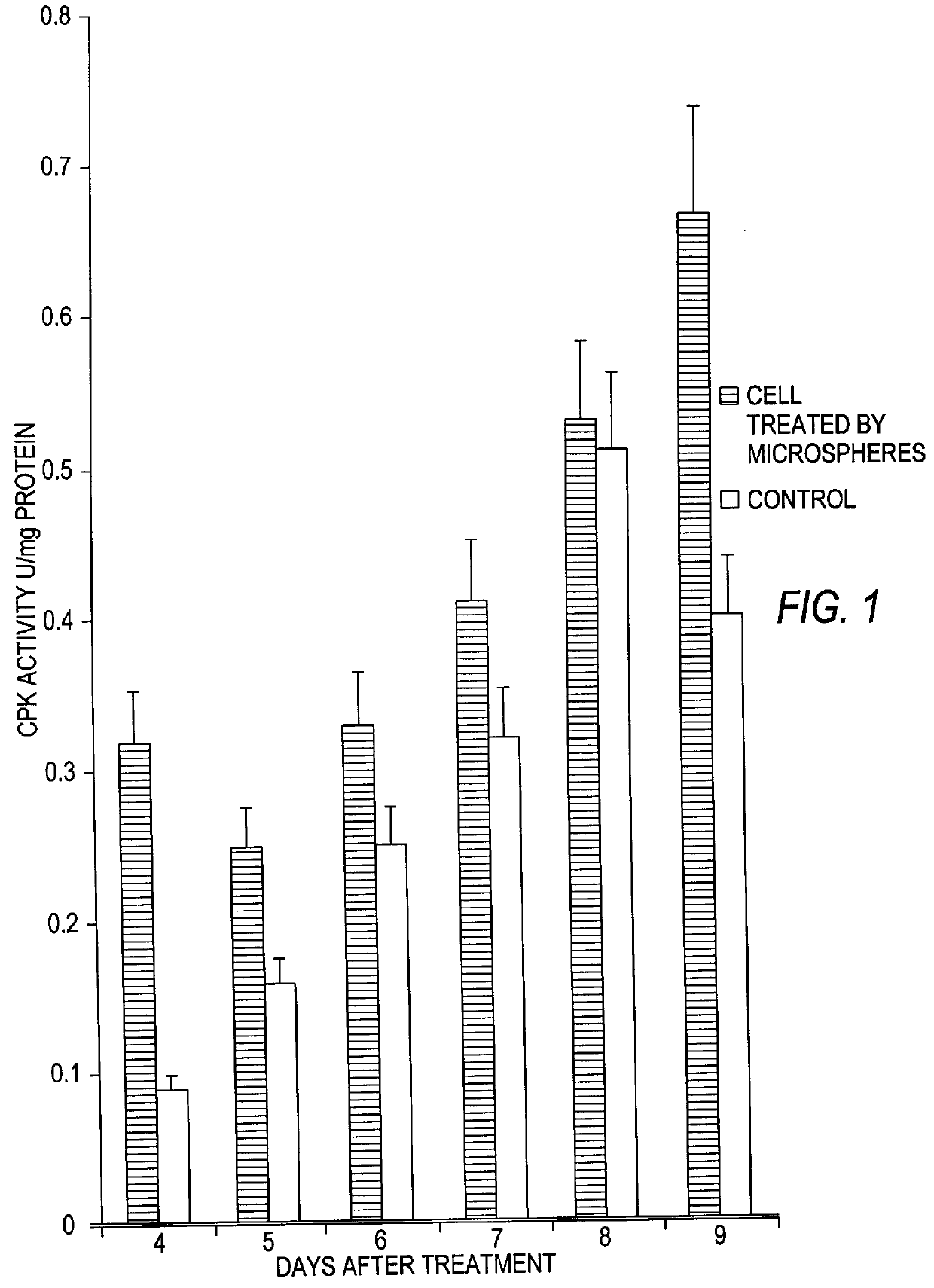
Effect of Microspheres on Creatine Phosphokinase
The microspheres of the present invention clearly induced an initial increase in creatine phosphokinase (CPK) activity of cultured myoblasts, as shown in FIG. 1. However, after eight days, the untreated and treated cells both demonstrate the same level of CPK activity, indicating that the induction of increased CPK activity by the microspheres of the present invention is temporary. The experimental method was as follows.
A primary culture of rat embryo skeletal muscle was prepared as described by Freshney [R. J. Freshney, Culture of Animal Cells, Willey. 1986, p. 117. 170-172]. Briefly, the muscles were dissected free of skin and bone and 15 desegregated by warm trypsinization (0.25% trypsin at 36.5.degree. C.). Contamination by fibroblasts was reduced by preplating cells for 1 hour in an incubator with 5% CO.sub.2, 37.degree. C., since fibroblasts adhere to tissue culture plates first. Myoblasts were then seeded on 35 mm Petri dishes a...
example 2
Effect of Microspheres on Cell Proliferation and Fusion
The microspheres of the present invention were demonstrated to induce an initial increase in both cell proliferation and myoblast fusion, as compared to control (untreated) cells, as shown below.
Primary cultures of rat myoblasts were prepared as described in Example 1 above, except that the cells were grown on cover slips. Treated cells were incubated with microspheres in media, as further described below, while control cells were only given media To determine the extent of cell proliferation, cells were fixed in ethanol / acetic acid (3:1) and then stained by hematoxilin-eosin. The stained cells were then counted in a light microscope. The mitotic index was calculated as the proportion of cells in mitosis counted per 1000 cells.
For the examination of cell proliferation, polystyrene microspheres which had sulfate surface groups were used, with a diameter of 0.18 microns, and a concentration of 10.sup.7 microspheres / ml of media. A ...
example 3
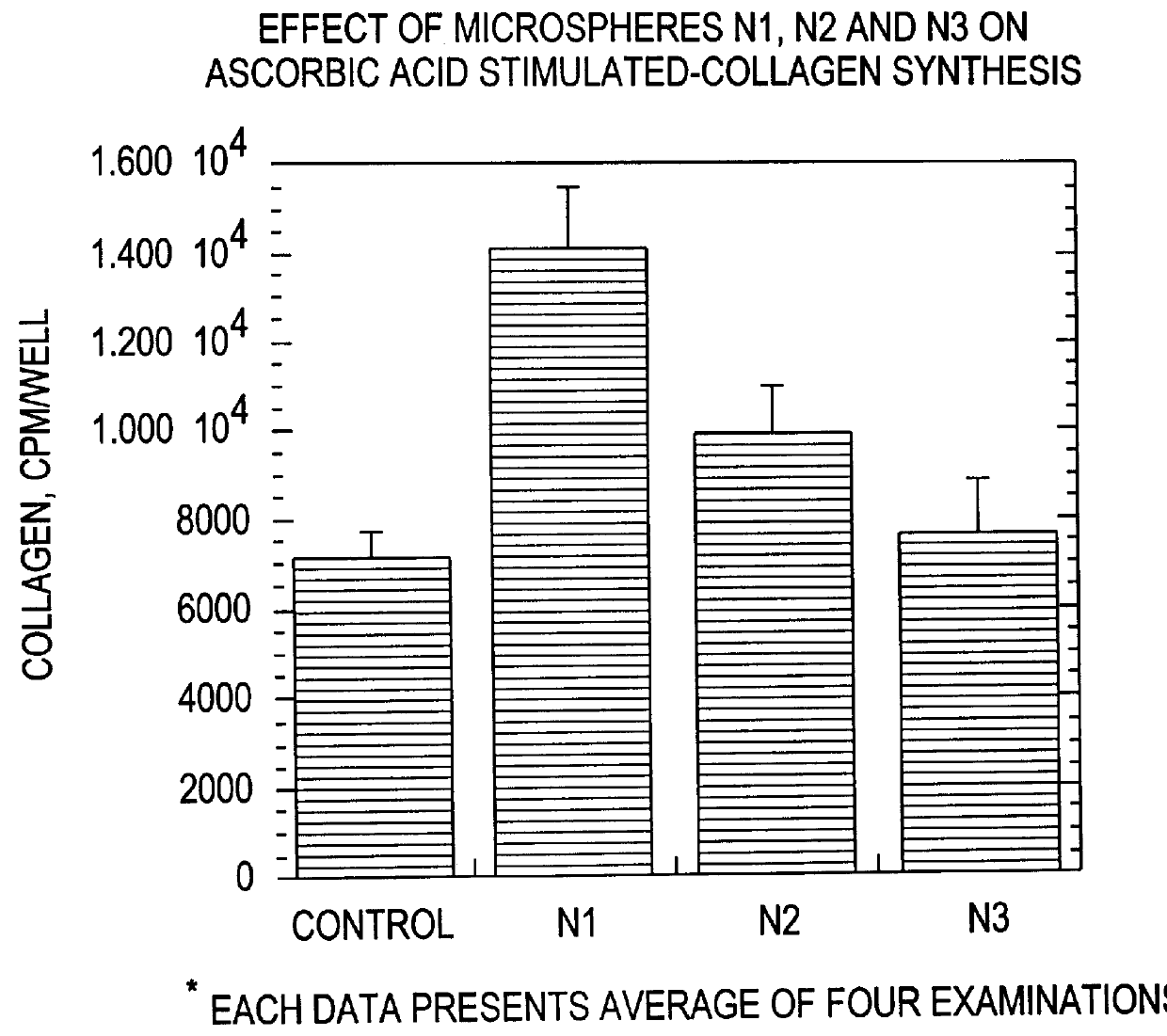
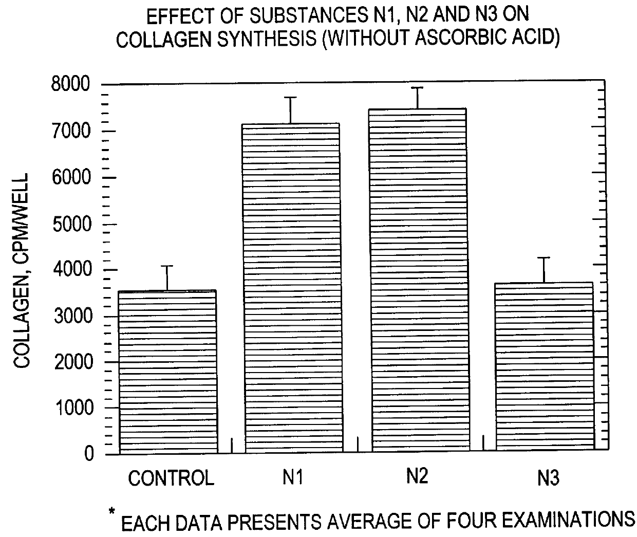
Effect of Microspheres on Collagen Synthesis and Deposition
As noted above in the Background section, collagen synthesis and deposition is an important step in the process of wound healing. Furthermore, the amount of collagen deposited in the !wound is an important determinant of wound strength. Thus, although the microspheres of the present invention clearly have a variety of effects on different cell types, as demonstrated in the preceding and following Examples, clearly one important determinant of the ability of a composition to promote wound healing is its effect on collagen synthesis and deposition.
As shown in FIGS. 2A and 2B, the microspheres of the present invention clearly promote collagen synthesis by cultured fibroblasts. The largest effect is seen with Type I and Type II microspheres. Type I microspheres had a diameter of 4.5 microns, was made of carboxylated polystyrene and had a Z potential of about -29.96 mV. Type II microspheres had a diameter of 0.49 microns, were ma...
PUM
| Property | Measurement | Unit |
|---|---|---|
| Angle | aaaaa | aaaaa |
| Angle | aaaaa | aaaaa |
Abstract
Description
Claims
Application Information