3d refractive index tomography and structured illumination microscopy system using wavefront shaper and method thereof

A wavefront shaper, structured light illumination technology, applied in microscopy, fluorescence/phosphorescence, coupling of optical waveguides, etc., can solve problems such as photobleaching

Active Publication Date: 2017-12-19
TOMOCUBE INC +1
View PDF9 Cites 3 Cited by
  • Summary
  • Abstract
  • Description
  • Claims
  • Application Information

AI Technical Summary

Problems solved by technology

However, in the case of fluorescent images, there are disadvantages in that the process of staining cells in order to find fluorescent proteins is infiltrating, and if the fluorescent images are measured for a long time, photobleaching occurs, making measurement impossible

Method used


Image

  • 3d refractive index tomography and structured illumination microscopy system using wavefront shaper and method thereof
  • 3d refractive index tomography and structured illumination microscopy system using wavefront shaper and method thereof
  • 3d refractive index tomography and structured illumination microscopy system using wavefront shaper and method thereof

Examples


Embodiment Construction

[0044] Hereinafter, embodiments will be described in detail with reference to the drawings. However, the described embodiments can be modified in various other forms, and the scope of the present invention is not limited to the embodiments described below. And, the various embodiments are provided in order to more fully explain the present invention to those having ordinary knowledge in the technical field. The shapes, sizes, and the like of elements in the drawings may be exaggerated for clearer illustration.

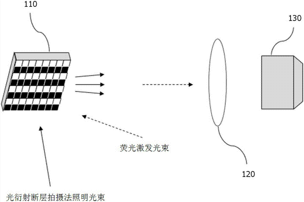
[0045] Grasping the internal structure of living cells three-dimensionally and measuring changes in the structure in real time is a technology that greatly contributes to biological and pathological research.

[0046] The following embodiments disclose a system and method capable of fully realizing 3D Refractive Index Tomogram and 3D Structured Illumination Microscopy by using a Wavefront Shaper.

[0047] The embodiment provides a technology for simultaneously measur...


PUM


Abstract

An ultra-high-speed 3D refractive index tomography and structured illumination microscopy system using a wavefront shaper and a method using the same are provided. A method of using an ultra-high-speed 3D refractive index tomography and structured illumination microscopy system that utilizes a wavefront shaper includes adjusting an irradiation angle of a plane wave incident on a sample by using the wavefront shaper, measuring a 2D optical field, which passes through the sample, based on the irradiation angle of the plane wave, and obtaining a 3D refractive index image from information of the measured 2D optical field by using an optical diffraction tomography or a filtered back projection algorithm.

Description

technical field [0001] The following embodiment relates to an ultra-high-speed three-dimensional refractive index image capture system using a wavefront shaper and a fluorescent structured light illumination microscope system and its application method. In more detail, it involves a method for simultaneously measuring the three-dimensional Technologies for high-resolution fluorescence images and three-dimensional refractive index stereograms. Background technique [0002] The current methods for optically measuring the internal structure of cells include the fluorescence image method for detecting fluorescent proteins in specific structures inside the cell and then measuring the fluorescent signal, and the three-dimensional refractive index tomography method for measuring the three-dimensional refractive index distribution inside the cell. [0003] Three-dimensional refractive index tomography, which measures the three-dimensional refractive index distribution inside the cel...

Claims


Application Information

Patent Timeline
Patent Type & Authority Applications(China)
IPC IPC(8): G01N21/45G01N21/64
CPCG01N21/455G01N21/6456G01N21/6486G01N2021/6495G01N21/45G01N21/6458G01N2021/1787G01N2201/0635G01N21/453G02B6/32G02B6/3516G01N21/4133G02B21/16G02B21/367G02B26/0833G02B27/46G03H1/0005G03H2001/005
Inventor 朴龙根申升雨朴光植
Owner TOMOCUBE INC
Why Eureka
Try Eureka