
Computational fluid mechanics-based cerebrovascular reserve capacity simulation system and method

A computational fluid dynamics and simulation system technology, applied in computing, radiological diagnostic instruments, medical science, etc., can solve problems such as invasive measurement methods, low spatial resolution, and inability to provide reserves of blood vessels and vascular networks , to avoid deviation

Active Publication Date: 2017-12-19
WUHAN UNIV
View PDF9 Cites 28 Cited by
  • Summary
  • Abstract
  • Description
  • Claims
  • Application Information

AI Technical Summary

Problems solved by technology

[0003] Traditional imaging techniques for evaluating cerebrovascular reserve after stroke mainly include PET imaging, SPECT imaging, Xenon-enhanced CT imaging, CT perfusion (Dynamic perfusion CT), MRI perfusion imaging (perfusion-weighted MRI), arterial Spin labeling imaging (Arterial Spin Labeling MRI), blood oxygen level-dependent imaging (Blood oxygen level-dependent MRI), etc., these technical means can provide local perfusion information in the region of interest of the brain, including hemodynamics such as CBF and CBV However, it cannot provide the specific reserve capacity of a single vessel, and the spatial resolution is low, and most of the above measurement methods are invasive and expensive
Transcranial Doppler Ultrasound and Phase-contrast MRI can non-invasively measure the flow velocity and flow data of local blood vessels, but Transcranial Doppler is easily affected by the operator. Influenced by subjective operations, and the two techniques can only provide information on a certain section of the vessel of interest at one time, but cannot provide the reserve force of the entire vessel and vascular network

Method used


Image

  • Computational fluid mechanics-based cerebrovascular reserve capacity simulation system and method
  • Computational fluid mechanics-based cerebrovascular reserve capacity simulation system and method
  • Computational fluid mechanics-based cerebrovascular reserve capacity simulation system and method

Examples


Embodiment Construction

[0031] In order to make the object, technical solution and advantages of the present invention more clear, the present invention will be further described in detail below in conjunction with the examples. It should be understood that the specific embodiments described here are only used to explain the present invention, not to limit the present invention.

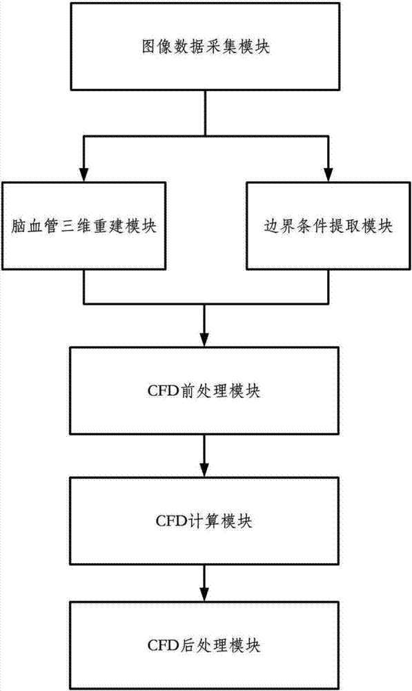
[0032] like figure 1 As shown, the cerebrovascular reserve force simulation system based on computational fluid dynamics provided in this embodiment includes,

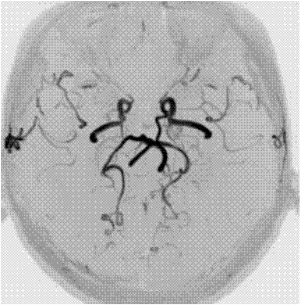
[0033] The image data acquisition module is used for acquiring computed tomography images of the geometric structure of cerebral vessels. In this example, the computed tomography images of head and neck vessels are collected on 3.0T magnetic resonance using TOF-MRA MRI sequence, as shown in figure 2 shown. The image data acquisition module also uses magnetic resonance Phase-contrast MRI (PC-MRI) phase-enhanced sequence to acquire phase-enhanced images of the ent...


PUM


Abstract

The invention discloses a computational fluid mechanics-based cerebrovascular reserve capacity simulation system and method. The system comprises an image data collection module used for obtaining a computed tomography image of a brain blood vessel of a human body, a blood vessel three-dimensional reconstruction module for performing three-dimensional reconstruction on the computed tomography image, a boundary condition extraction module for performing post-processing on the computed tomography image of the brain blood vessel to obtain boundary information required for simulation, a CFD preprocessing module for performing preprocessing required for numerical simulation on a three-dimensional geometric model of the brain blood vessel, a CFD computing module for performing solving on hemodynamics information of all parts of the three-dimensional geometric model of the brain blood vessel, and a CFD post-processing module for comparing a simulation result with a result measured by the boundary condition extraction module. Hemodynamic feature parameters are adopted as standards for evaluating cerebrovascular reserve capacity, so that the one-sidedness of judgment through a geometric structure of the brain blood vessel is avoided, and noninvasive, quantitative and personalized cerebrovascular reserve capacity evaluation can be realized.

Description

technical field [0001] The invention relates to medical device technology, in particular to a cerebrovascular reserve force simulation system and method based on computational fluid dynamics. Background technique [0002] Stroke, commonly known as stroke, is a general term for a class of acute cerebrovascular diseases, manifested as acute and chronic cerebral hemorrhage or ischemic symptoms. It has the characteristics of high morbidity, high mortality and high disability rate, and it is the primary factor leading to the death and disability of adults in our country. Relevant clinical studies have found that based on imaging, the evaluation of cerebrovascular reserve plays a very important role in the prevention, diagnosis and treatment of stroke. [0003] Traditional imaging techniques for evaluating cerebrovascular reserve after stroke mainly include PET imaging, SPECT imaging, Xenon-enhanced CT imaging, CT perfusion (Dynamic perfusion CT), MRI perfusion imaging (perfusion...

Claims


Application Information

Patent Timeline
Patent Type & Authority Applications(China)
IPC IPC(8): G06F19/00G06T17/00A61B6/00A61B6/03
CPCA61B6/03A61B6/507A61B6/5205A61B6/5229G06T17/00
Inventor 徐海波孙文博阮兆方键孙振萌
Owner WUHAN UNIV
Learn More