Method for preparing anti-EGFR safe chimeric antigen receptor modified immune cell and application thereof

A chimeric antigen receptor and immune cell technology, applied in the field of immune cell preparation, can solve the problems of false attack of tissue cells, cytokine storm, etc., and achieve the effects of controlling toxic side effects, improving safety, and avoiding adverse reactions

Active Publication Date: 2018-01-09
山东省成体细胞产业技术研究院有限公司
View PDF4 Cites 4 Cited by
  • Summary
  • Abstract
  • Description
  • Claims
  • Application Information

AI Technical Summary

Problems solved by technology

However, in clinical applications, CAR-T cells may accidentally attack normal tissue cells, or cause toxic side effects such as cytokine storm due to the massive release of inflammatory cytokines.

Method used


Image

  • Method for preparing anti-EGFR safe chimeric antigen receptor modified immune cell and application thereof
  • Method for preparing anti-EGFR safe chimeric antigen receptor modified immune cell and application thereof
  • Method for preparing anti-EGFR safe chimeric antigen receptor modified immune cell and application thereof

Examples


Embodiment 1

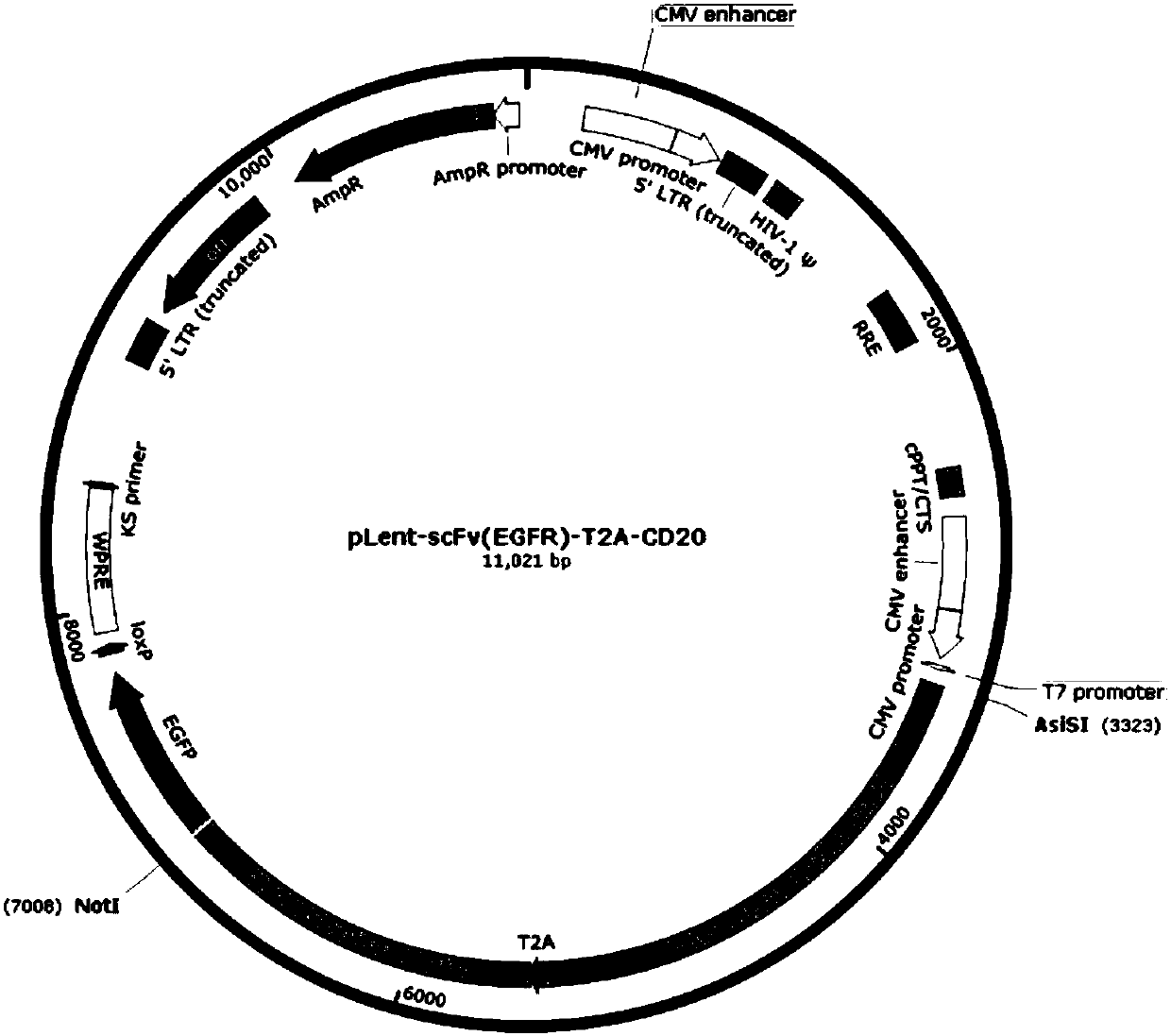
[0025] Embodiment 1: the fusion gene fragment scFv (EGFR)-CD8-CD137-CD3ζ-T2A-Leader-CD20-CD8-CD3ζ is inserted into the lentiviral expression vector pLent-C-GFP

[0026] Anti-EGFR CAR module comments figure 1 (See appendix SEQ ID NO.1 for the nucleic acid sequence).

[0027] The sequence of each module of CAR of Anti-EGFR

[0028] (1) CD20 nucleic acid artificial sequence (SEQ ID NO.2)

[0029] (2) Linker nucleic acid artificial sequence (SEQ ID NO.3)

[0030] (3) T2A nucleic acid artificial sequence (SEQ ID NO.4)

[0031] (4) CAR leader sequence (Leader) nucleic acid artificial sequence (SEQ ID NO.5)

[0032] (5) Anti-EGFR single chain Fv antibody nucleic acid artificial sequence (SEQ ID NO.6)

[0033] (6) CD8 Hinge region nucleic acid artificial sequence (SEQ ID NO.7)

[0034] (7) CD8 transmembrane region nucleic acid artificial sequence (SEQ ID NO.8)

[0035] (8) CD137 intracellular nucleic acid artificial sequence (SEQ ID NO.9)

[0036] (9) CD3ζ intracellular region...

Embodiment 2

[0038] Embodiment 2: lentivirus packaging, virus titer detection

[0039] Inoculate the cell line 293T in a 10 cm dish containing DMEM+10% FBS at 37°C, 5% CO 2 Cultured under conditions, the adherence rate is 70%-80%, and then used for lentivirus transfection. Recombinant plasmid pLent-scFv(EGFR)-T2A-CD20 and empty plasmid pLent-C-GFP were co-transfected with lentiviral packaging plasmid into 293T cells by calcium phosphate transfection method. For specific methods, refer to molecular cloning. 24 hours after transfection, the cells were obviously enlarged and spherical, and their ability to adhere to the wall decreased and they were easy to fall off. After 48 hours, the expression of green fluorescent protein in the cells was observed under an inverted fluorescence microscope. After 72 hours, collect the supernatant into an EP tube, centrifuge at 2000 g for 10 min, transfer to a new EP tube, filter through a 4.5 μm filter, and store the virus solution at -80°C. According to...

Embodiment 3

[0040] Example 3 Lentivirus Infection of CIK Cells and Expansion and Culture of CIK Cells after Infection

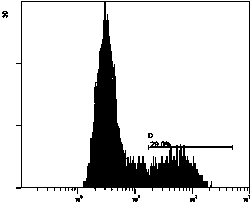
[0041] Infect CIK cells with the above-mentioned recombinant lentivirus, incubate the infected cells in a 37°C, 5% CO2 incubator for 8 hours, collect the cells, add virus solution again, 1000g, centrifuge for 90 minutes, and culture at 37°C, 5% CO2 Continue to culture in the box, so repeated multiple infection, improve the infection efficiency of CIK cells. Aspirate 2ml of the culture supernatant, add 2ml of fresh medium, continue to expand the culture, and culture for 17 days until the cells are expanded to a sufficient amount. The chimeric antigen receptor expression was detected by the FLTC (isothiocyanate) channel of the FC500 flow cytometer. Taking uninfected CIK lymphocytes as a negative control, the positive rate of CIK cells infected by recombinant lentivirus was 29.0% ( image 3 ).


PUM


Abstract

The invention discloses a method for preparing an anti-EGFR safe chimeric antigen receptor modified immune cell and application thereof. The method comprises the following steps: synthesizing scFv(EGFR)-CD8-CD137-CD33 zeta-T2A-Leader-CD20-CD8-CD3 zeta-nucleotide; inserting a fusion gene fragment into a lentivirus expression vector, and packaging into a lentivirus carrying scFv(EGFR)-CD8-CD137-CD3zeta-T2A-Leader-CD20-CD8-CD3 zeta coding genes, and infecting a monocyte induced immune cell to obtain the anti-EGFR safe chimeric antigen receptor modified immune cell. According to the method, the safety of chimeric antigen receptor modified immune cell can be improved, toxic and side effect can be controlled, and occurrence of adverse response can be avoided.

Description

technical field [0001] The invention relates to the field of biological genes, more specifically, a preparation method and application of an anti-EGFR safe chimeric antigen receptor modified immune cell. Background technique [0002] EGFR (epidermal growth factor receptor) is the expression product of the proto-oncogene c-erbB1, and is a member of the epidermal growth factor receptor (HER) family. As a cell surface protein, EGFR is expressed on the surface of normal epithelial cells, but is expressed at abnormally high levels on the surface of many types of cancer cells, such as kidney cancer, lung cancer, prostate cancer, pancreatic cancer, breast cancer and other tissues. EGFR is related to tumor cell proliferation, angiogenesis, tumor invasion, metastasis and inhibition of apoptosis. It can be seen that EGFR is closely related to a variety of cancers, and is one of the ideal targets for tumor targeted therapy and diagnosis. [0003] Chimeric antigen receptor T cells (ch...

Claims


Application Information

Patent Timeline
Patent Type & Authority Applications(China)
IPC IPC(8): C12N15/867C12N15/62C12N5/10A61K35/17A61P35/00
Inventor 刘明录王立新韩庆梅王亮万磊金海锋刘敏张传鹏强邦明
Owner 山东省成体细胞产业技术研究院有限公司
Why Eureka
Try Eureka