
Method and system for detection based on brain medical imaging

A medical imaging and detection method technology, applied in the medical field, can solve the problems of image brightness sensitivity, inability to effectively use the position information of image features, brightness insensitivity, etc., and achieve the effect of a wide range of clinical applications

Active Publication Date: 2018-01-16
陈烨 +1
View PDF8 Cites 4 Cited by
  • Summary
  • Abstract
  • Description
  • Claims
  • Application Information

AI Technical Summary

Problems solved by technology

[0010] Traditional algorithms can effectively process the location information of image features, but they are sensitive to image brightness. FBM is not sensitive to image brightness, but cannot effectively use the location information of image features.

Method used


Image

  • Method and system for detection based on brain medical imaging
  • Method and system for detection based on brain medical imaging
  • Method and system for detection based on brain medical imaging

Examples


Embodiment Construction

[0063] In order to make the object, technical solution and advantages of the present invention more clear, the present invention will be further described in detail below in conjunction with the examples. It should be understood that the specific embodiments described here are only used to explain the present invention, not to limit the present invention.

[0064] The application principle of the present invention will be further described below in conjunction with the accompanying drawings and specific embodiments.

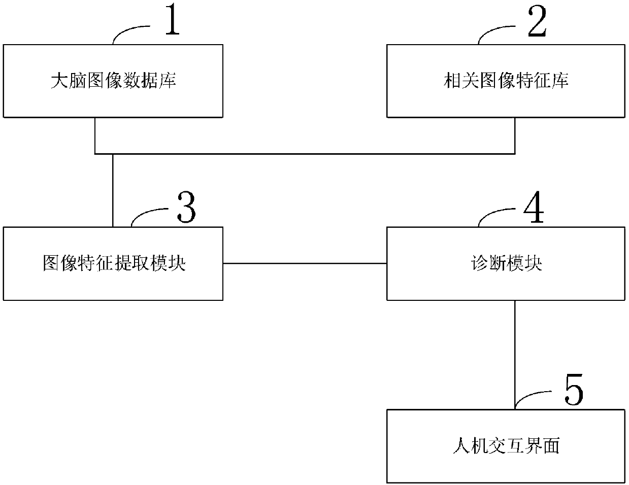
[0065] Such as figure 1 As shown, the detection system based on brain medical imaging provided by the embodiment of the present invention includes:

[0066] Brain image database 1, used to store brain medical image data of brain disease patients and non-patients, as the basis for extracting disease-related image features;

[0067] Related image feature library 2, used to save image features of lesions in various regions of the brain;

[0068] Image feature ext...


PUM


Abstract

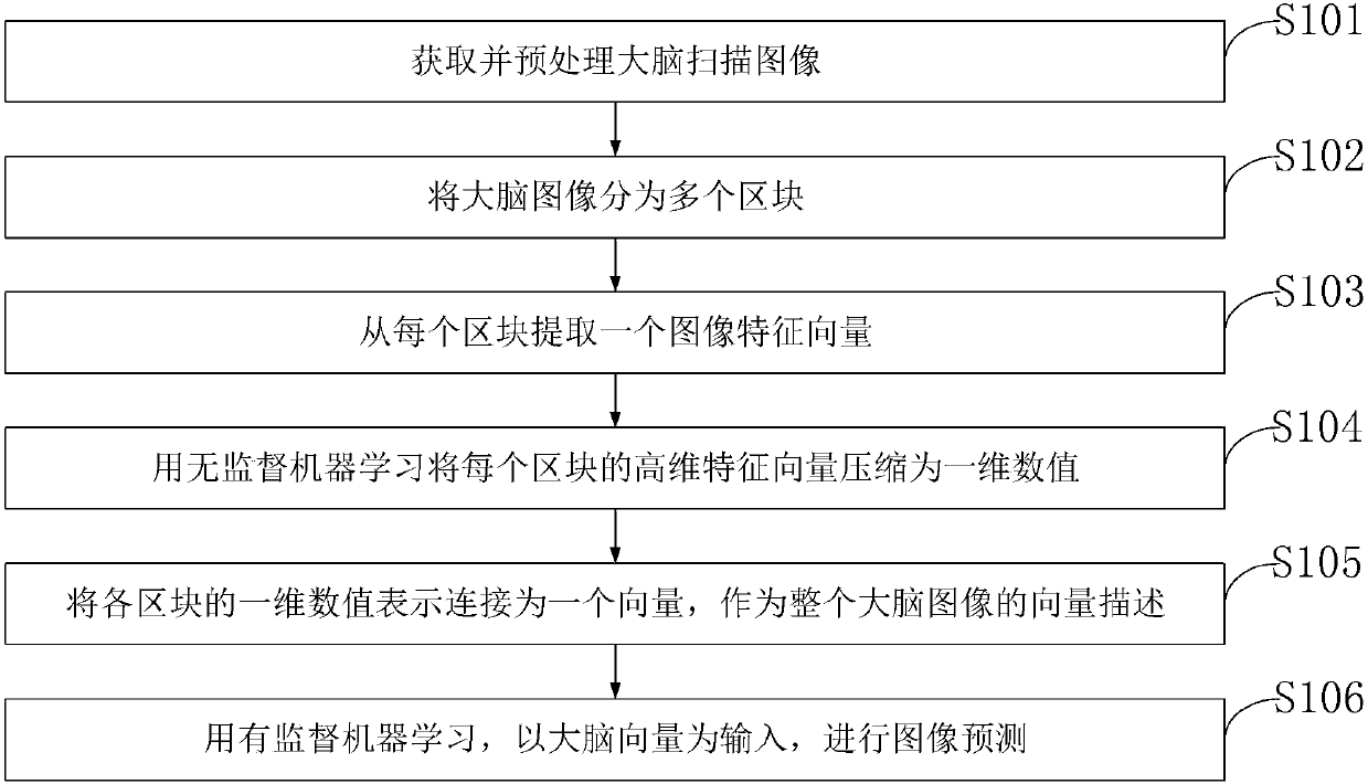
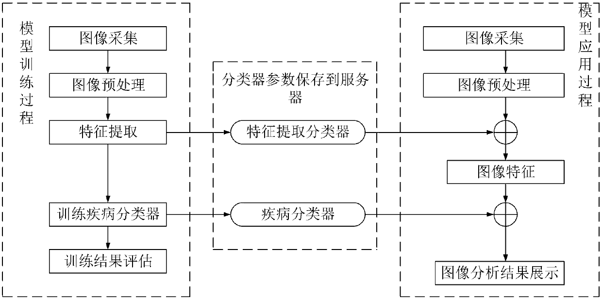
The present invention belongs to the medical technology field, and discloses a method and system for detection based on brain medical imaging. A machine learning algorithm is employed to automaticallyextract graphic features of each region of a brain; and using machine learning to judge a diagnosis result based on the extracted image features. The detection system comprises: a brain image database, a related image feature library, an image feature extraction module, a diagnostic module and a human-computer interaction interface. According to the method for detection based on brain medical imaging provided in the embodiment of the present invention, an image feature automatic extraction algorithm uses a machine learning algorithm to automatically extract the graphic features of various regions of the brain; and the diagnostic algorithm uses the machine learning to provide diagnostic results and diagnostic evidenced based on the image features.

Description

technical field [0001] The invention belongs to the field of medical technology, in particular to a detection method and system based on brain medical imaging. Background technique [0002] Current clinical diagnoses of brain disorders are often complemented by non-invasive imaging of the brain. However, the analysis and diagnosis of brain images requires a lot of time and effort of relevant disease experts, and the diagnosis results are easily affected by subjective factors. First of all, the differences in brain images caused by brain diseases are usually small and only exist in one or more specific positions of the images. Therefore, it takes a lot of time to find these lesion positions from 3D brain images. Secondly, there are individual differences in the structure of the brain, and it is also a difficult problem to accurately distinguish between individual differences and differences caused by lesions, which are easily affected by the personal experience of experts an...

Claims


Application Information

Patent Timeline
IPC IPC(8): G06T7/00G06T3/00
Inventor 陈烨卢龙
Owner 陈烨
Learn More