
Super-thick tissue slice preparation method and method for reproducing three-dimensional shape structure at a cell level

A tissue section and cell-level technology, which can be used in the field of reproducing the three-dimensional morphological structure of tissue at the cell level and the preparation of ultra-thick tissue sections. Effect

Active Publication Date: 2018-02-23
ZHUJIANG HOSPITAL SOUTHERN MEDICAL UNIV
View PDF3 Cites 12 Cited by
  • Summary
  • Abstract
  • Description
  • Claims
  • Application Information

AI Technical Summary

Problems solved by technology

Using thick tissue slices (100 μm) to obtain 3D structural images of the liver is limited by the penetration depth of laser light and the penetration of antibodies, and the observation thickness is ≤100 μm. Therefore, the imaging depth of the Z axis is still smaller than the diameter of a single hepatic lobule, which cannot be achieved. ideal viewing depth

Method used


Image

  • Super-thick tissue slice preparation method and method for reproducing three-dimensional shape structure at a cell level
  • Super-thick tissue slice preparation method and method for reproducing three-dimensional shape structure at a cell level
  • Super-thick tissue slice preparation method and method for reproducing three-dimensional shape structure at a cell level

Examples


Embodiment Construction

[0056] The present invention will be described in further detail below in conjunction with the accompanying drawings and specific embodiments.

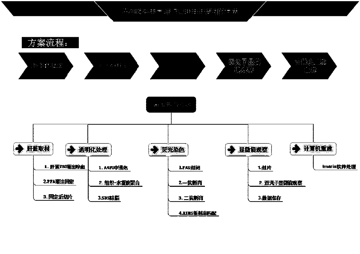
[0057] In this example, relevant reagents are used to remove lipids in the liver and other substances that cannot penetrate light, so as to realize the transparency of the liver. At the same time, the tissue block is directly soaked in the antibody for incubation to improve the depth of immunostaining. Specifically, refer to figure 1 , including the following steps:

[0058] 1. Obtain the tissue material to be observed.

[0059] The tissue material is derived from a fresh organ or tissue. Usually, these fresh organs or tissues prepared for tissue sectioning require at least one of the following pretreatments:

[0060] (1) Cleaning the surface of the fresh organ or tissue to remove blood stains on the surface, parts of other organs or tissues that do not belong to the organ or tissue, or parts detached from the organ or tissue;

[0...


PUM


Abstract

The present invention discloses a super-thick tissue slice preparation method, which comprises: obtaining a tissue material to be observed; preparing tissue material slices with a thickness of 1-8 mm,and carrying out transparentizing on the tissue material slices; and carrying out fluorescent staining on the transparent tissue material slices. The method for reproducing the three-dimensional tissue structure of a tissue slice comprises: preparing a transparent tissue slice with a thickness of 1-8 mm; obtaining the image data of the transparent tissue slice by using a fluorescence microscope;recombining the image data to form three-dimensional model data; and based on the three-dimensional model data, outputting a three-dimensional image. According to the present invention, with the super-thick tissue slice preparation method and the three-dimensional tissue structure reproducing method, the tissue slice is subjected to grease removing to achieve the transparent state so as to increase the penetrating depth of laser in liver tissue and reduce the light scattering, such that the deep imaging of the tissue can be achieved, the real cell structure of the tissue can be reflected, andthe imaging efficiency and the fidelity can be improved.

Description

technical field [0001] The invention relates to the field of microscopic observation of biological tissues, in particular to a method for preparing ultra-thick tissue slices and reproducing the three-dimensional morphological structure of tissues at the cell level. Background technique [0002] When analyzing the detailed information of cells or subcellular structures of organs or tissues, 3-10 μm 2D tissue slices are a routine method to achieve this requirement. Usually, the organ / tissue is fixed, embedded in paraffin, sectioned (or frozen section), and then stained accordingly and observed under a microscope. This method can morphologically analyze the various cell types and their distribution in the organ / tissue Research. However, this method is only limited to a certain plane level and does not necessarily include the whole organ / tissue, especially in the pathological analysis of the organ, taking the liver as an example, the existing technology cannot include the liver...

Claims


Application Information

Patent Timeline
IPC IPC(8): G01N1/28G01N1/30G01N1/34G01N33/533G01N21/64
CPCG01N1/28G01N1/286G01N1/30G01N1/34G01N21/6402G01N21/6458G01N21/6486G01N33/533G01N2001/2873G01N2001/305
Inventor 汪艳郑永见潘明新李阳
Owner ZHUJIANG HOSPITAL SOUTHERN MEDICAL UNIV
Learn More