

Fluorescence detection kit for simultaneous detection of three kinds of breast cancer tumor markers and detection method thereof
A breast cancer tumor and fluorescence detection technology is applied to a fluorescence detection kit and detection field for simultaneous detection of three breast cancer tumor markers, which can solve the problems of long detection time, weak specificity, large errors, etc.
- Summary
- Abstract
- Description
- Claims
- Application Information
AI Technical Summary
Problems solved by technology
Method used
Image

Examples
Embodiment 1
[0043] A fluorescent detection kit for the simultaneous detection of three breast cancer tumor markers, the three breast cancer tumor markers are respectively CEA, CA153, and CA125, and the kit includes 10 solutions, which are respectively G1 solution, G2 liquid, G3 liquid, Q1 liquid, Q2 liquid, Q3 liquid;
[0044] The G1 solution is a graphene oxide solution labeled with CEA antigen, the concentration is 5 μgmL -1 ; The G2 solution is a graphene oxide solution labeled with the CA153 antigen, with a concentration of 10 μg mL -1 ; G3 solution is a graphene oxide solution labeled with CA125 antigen, the concentration is 100μgmL -1; The three kinds of graphene oxide surfaces have carboxyl groups, and the carboxyl groups are coupled with corresponding CEA antigens, CA153 antigens, and CA125 antigens respectively;
[0045] The Q1 solution is a fluorescent quantum dot solution labeled with the CEA antibody, the concentration is 1 μM, and the maximum emission wavelength is 620 nm; ...
Embodiment 2
[0057] The parts of the kit are the same as in Example 1. The method for detecting the breast cancer tumor marker CA153 using the kit above,
[0058] Step 1: Mix Q1 liquid, Q2 liquid, and Q3 liquid according to the volume ratio of 1:1:1 to obtain A mixed liquid; mix G1 liquid, G2 liquid, and G3 liquid according to the volume ratio of 1:1:1, Obtain B mixed solution;
[0059] Step 2: Dilute S2 with H solution, and add 1, 10, 100, 1000, 10000nU mL in a 96-well plate -1 S2 15 μL;
[0060] Step 3: Then add 15 μL of mixed solution A and 60 μL of mixed solution B;
[0061] Step 4: Carry out incubation, the temperature is 37 degrees Celsius, and the incubation time is 120 minutes;
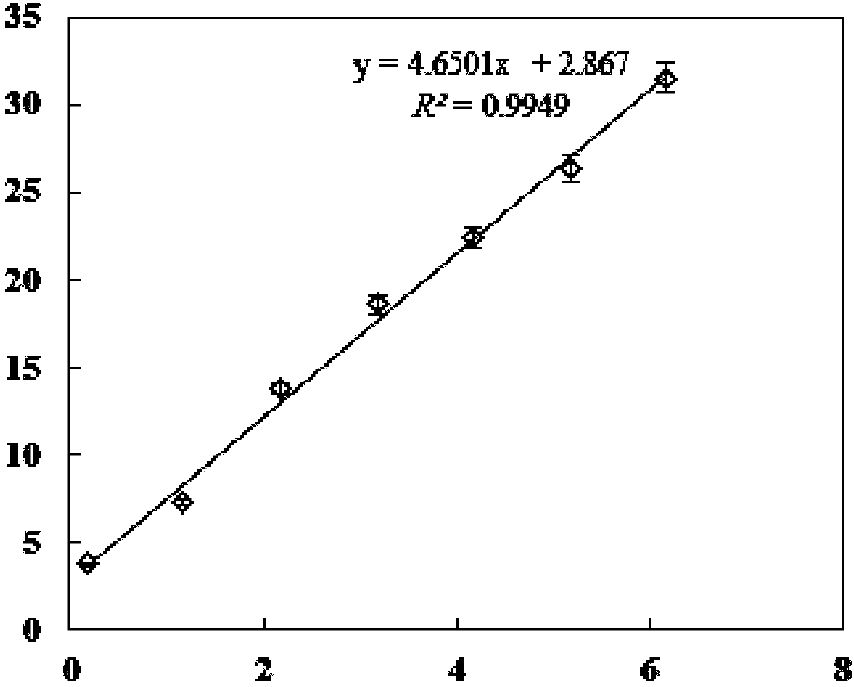
[0062] Step 5: Put the mixed solution into a microplate reader to detect the fluorescence signal, the fluorescence excitation wavelength should be below 400nm, and measure the fluorescence intensity of each well at 460nm, 540nm and 619nm respectively;
[0063] Step 6: The linear range between the blan...
Embodiment 3
[0066] The parts of the kit are the same as in Example 1. Utilize above-mentioned kit to carry out the method for breast cancer tumor marker CA125 detection,
[0067] Step 1: Mix Q1 liquid, Q2 liquid, and Q3 liquid according to the volume ratio of 1:1:1 to obtain A mixed liquid; mix G1 liquid, G2 liquid, and G3 liquid according to the volume ratio of 1:1:1, Obtain B mixed solution;
[0068] Step 2: Dilute S3 with H solution, and add 1, 5, 10, 100, 1000μUmL in 96 microwell plates -1 S3 15 μL;
[0069] Step 3: Then add 15 μL of mixed solution A and 60 μL of mixed solution B;
[0070] Step 4: Carry out incubation, the temperature is 37 degrees Celsius, and the incubation time is 120 minutes;
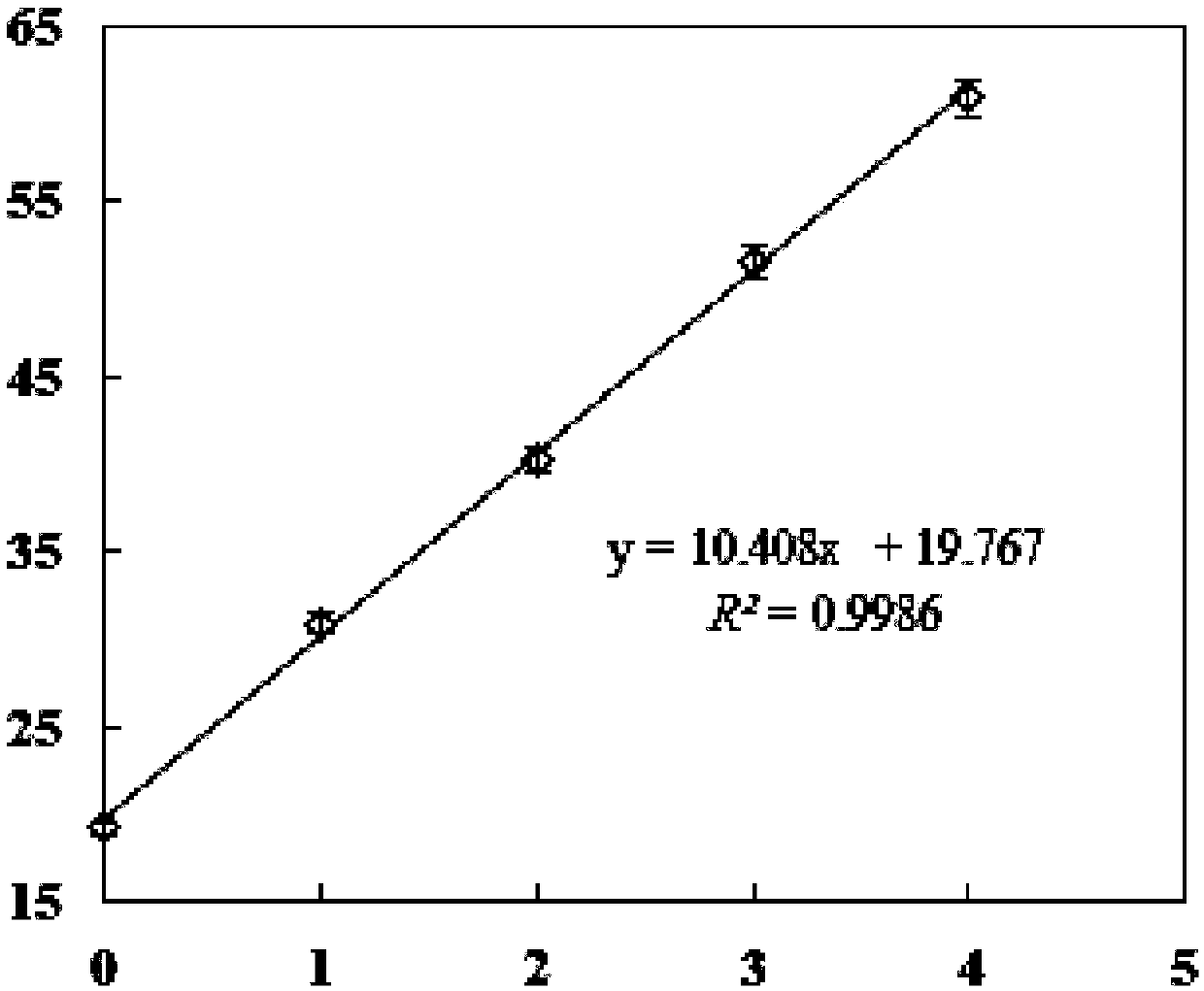
[0071] Step 5: Put the mixed solution into a microplate reader to detect the fluorescence signal, the fluorescence excitation wavelength should be below 400nm, and measure the fluorescence intensity of each well at 460nm, 540nm and 619nm respectively;
[0072] Step 6: The linear range ...
PUM
 Abstract
Description
Claims
Application Information