An image processing method of ultra-thin flexible electronic endoscope

An electronic endoscope and image processing technology, applied in image communication, television, electrical components, etc., can solve problems such as interference, patient bladder spasm, fixed pattern noise, dark current noise, and large thermal noise, and achieve good economic and social benefits, improved image quality, and high-resolution effects

Active Publication Date: 2022-01-28
上海瑞烁信息科技有限公司
View PDF7 Cites 0 Cited by
  • Summary
  • Abstract
  • Description
  • Claims
  • Application Information

AI Technical Summary

Problems solved by technology

When inserting from the urethra, the damage to the surrounding tissue is relatively large, especially for male patients, the urethra is relatively long (18-20cm), and the insertion has to go through two physiological bends and the external urethral sphincter, which is especially painful. Pain and bleeding, and in some patients bladder spasms, making examination difficult
Because it cannot be bent, there is a blind spot during the inspection, and it is easy to miss important lesions
[0004] At present, most endoscopes on the market use large-size CMOS chips, and the processing solution is to use DSP control chips for video output processing. When the noise of the CMOS video chip is small, the image quality is OK; but when the CMOS video chip is very In small cases, it is impossible to achieve good image quality by using a simple DSP control circuit. It is necessary to use a dedicated image processing chip and image software processing to achieve better results.
[0005] The emergence of flexible electronic cystoscopes has solved the above problems. At present, the size of flexible electronic cystoscopes at home and abroad is generally above 3mm. Therefore, the small-size application market is basically occupied by fiberscopes, and there is a technical defect that the product size is too large; For ultra-small sensors, considering the process design, the image source distance is very small, so the electrical interference generated leads to very large fixed pattern noise FPN (Fixed pattern noise), dark current noise, and thermal noise of the output original image data.

Method used


Image

  • An image processing method of ultra-thin flexible electronic endoscope

Examples


Embodiment Construction

[0024] The present invention will be further described below with reference to the accompanying drawings, but is not as limiting of the present invention:

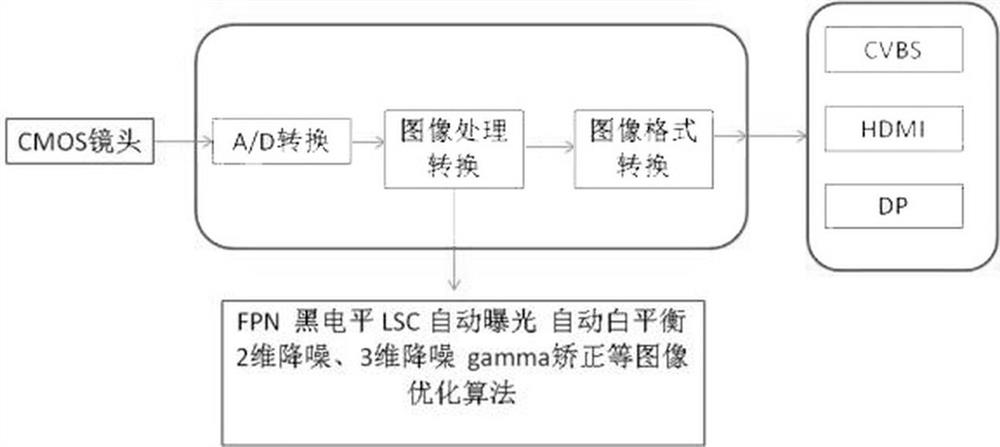
[0025] A ultrafine soft electron endoscope image processing method, including an image processing chip, the image processing chip consists of an optical imaging module, an AD conversion module, an image processing conversion module, and an image format conversion module, and an optical imaging module is CMOS. The lens, the image processing conversion module is the image-in-cell AD processing module, the image format conversion module is a real-time image processing system; includes the following steps:

[0026] A) The external cold light source passes through the fiber optic light to irradinate the surface of the organ mucosa;

[0027] B), the micro-optical lens at the front end of the CMOS lens aggregates the light reflected by the mucosa and projected to the photosensitive surface of the CMOS image sensor, and the CMOS senso...


PUM


Abstract

A method for image processing of an ultra-thin and flexible electronic endoscope, comprising the following steps: A), an external cold light source guides light through an optical fiber to the surface of the visceral mucosa in the body; B), the micro-optical lens at the front end of the CMOS lens gathers the mucosa The reflected light is projected onto the photosensitive surface of the CMOS image sensor, and the image data is obtained through the CMOS sensor configured by the controller; C), the image data is converted into a serial analog signal data stream by the high-precision code conversion chip of the AD conversion module; D ), the encoded image data is transmitted to the image reverse encoding AD processing module; E), the image reverse encoding AD processing module decodes the encoded analog signal, and inputs it into the real-time image processing system for processing; F), through the video encoder Video encoding and format conversion and output to the monitor to display clear and stable endoscopic images. The invention realizes long-distance high-fidelity transmission of image data and greatly improves image quality.

Description

Technical field [0001] The present invention belongs to the technical field of medical equipment, and in particular, the present invention relates to an ultrafine soft electron endoscope image processing method. Background technique [0002] The development of endoscopes has exceeded 200 years, experienced hard endoscopy, fiber optic endoscope, electron endoscope, capsule endoscope development stage, well known ultrafine diameter endoscope will be in medical minimally invasive / non-invasive , Industrial precision non-destructive testing, micro traces of public inspection system has broad application prospects, my country's "National Key R & D Plan: Basic Material Technology Improvement and Industrialization Key Special 2016 Project Declaration Guide" included in ultrafine diameter endoscope The development direction will play an important role in the next decade. [0003] Cystoscopy is one of the most basic examination methods of urology, and it is in the past, and there is a ha...

Claims


Application Information

Patent Timeline
Patent Type & Authority Patents(China)
IPC IPC(8): H04N5/225H04N5/374H04N5/378H04N5/357H04N7/18H04N7/01
CPCH04N7/01H04N7/18H04N23/555H04N23/56H04N23/54H04N23/55H04N25/60H04N25/76H04N25/75
Inventor 彭剑
Owner 上海瑞烁信息科技有限公司
Why Eureka
Try Eureka