
A group of associated proteins and their applications for assessing the prognosis of glioblastoma

A glioblastoma and protein technology, applied in the field of medical biological detection, to achieve the effect of improving sensitivity and improving survival rate

Inactive Publication Date: 2020-08-07
SECOND AFFILIATED HOSPITAL SECOND MILITARY MEDICAL UNIV
View PDF1 Cites 0 Cited by
  • Summary
  • Abstract
  • Description
  • Claims
  • Application Information

AI Technical Summary

Problems solved by technology

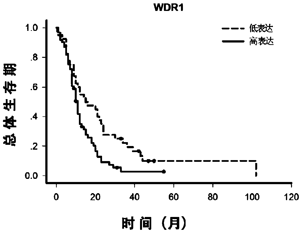
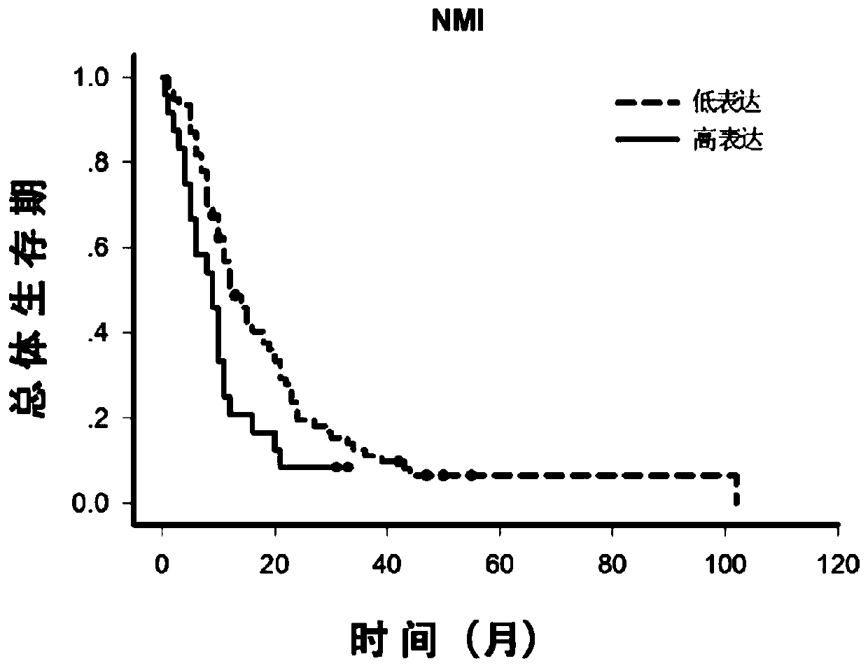
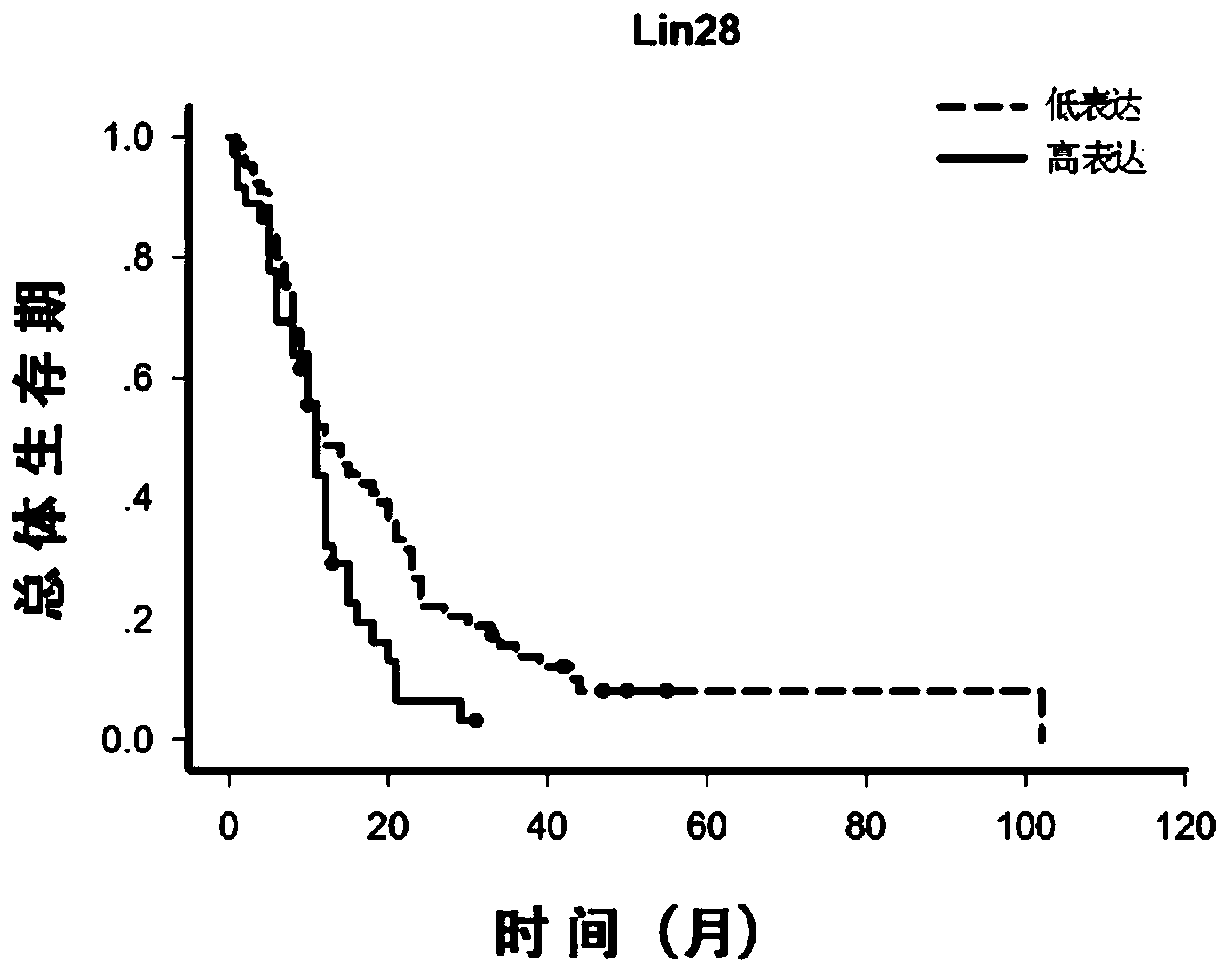
[0010] At present, there are no literature reports on the application of WDR1, NMI, and LIN28 proteins in glioblastoma

Method used


Image

  • A group of associated proteins and their applications for assessing the prognosis of glioblastoma
  • A group of associated proteins and their applications for assessing the prognosis of glioblastoma
  • A group of associated proteins and their applications for assessing the prognosis of glioblastoma

Examples


Embodiment 1

[0047] Example 1: Fabrication of Tissue Chips

[0048] 1. Main instruments and equipment

[0049] The main equipment includes automatic tissue dehydrator (Lycra ASP300), automatic paraffin embedding machine (Lycra

[0050] EG1160), automatic paraffin slicer (Leica RM2165), tissue chip microarray spotting instrument (Shanghai Bonan Biology), automatic staining machine (Meikang HMS740).

[0051] 2. Specific steps:

[0052] (1) Select a representative tissue sample, fix it in 10% neutral formalin, and divide the sample into 5mm×15mm×15mm;

[0053] (2) Use a fully automatic tissue dehydrator to dehydrate tissue samples;

[0054] (3) Use a paraffin embedding machine to embed the specimen into a paraffin block;

[0055] (4) Use a fully automatic paraffin slicer to slice the paraffin block into 4 microns and then slice the pathological tissue;

[0056] (5) Routine HE staining of pathological tissue sections using a fully automatic staining agent;

[0057] (6) Chip micro-display...

Embodiment 2

[0067] Example 2: Immunohistochemical staining

[0068] Main reagents include ethanol, xylene, H2O2, methanol, PBS (phosphate buffer saline), citrate buffer, 1% BSA blocking solution, DAB chromogen, hematoxylin, horseradish peroxidase; WDR1 antibody, NMI antibody and LIN28 antibody. Proceed as follows.

[0069] 1. Preparation of the tissue chip before use

[0070] 1) The tissue chip sealed in paraffin was baked at 60°C for 3 hours before use to melt the sealing wax on the surface;

[0071] 2) Soak the tissue chip in xylene for 10 minutes, repeat 4 times;

[0072] 3) Soak the tissue chip in absolute ethanol for 5 minutes, repeat twice; soak in 95% ethanol for 5 minutes; soak in 85% ethanol for 5 minutes; soak in 70% ethanol for 5 minutes; soak in distilled water for 5 minutes;

[0073] 2. Antigen heat recovery

[0074] Heat and boil 0.01M citrate buffer, place the tissue chip in the boiling buffer, continue to heat and pressurize for 5 minutes, cool to room temperature, ta...


PUM


Abstract

The invention relates to the technical field of medical biological detection and provides novel uses of WDR1, NMI and LIN28 proteins, particularly an application in preparation of a kit for assessingbrain glioblastoma prognosis. The invention further provides a kit and detection method performing brain glioblastoma prognosis assessing through quantitative detection of the WDR1, NMI and LIN28 proteins by adopting an immunohistochemical method. Expression levels of the WDR1, NMI and LIN28 proteins in brain glioblastoma tissues are measured by utilizing an immunohistochemical technique and system scoring, and it is determined that the expression levels of the WDR1, NMI and LIN28 proteins are related with prognosis of patients with brain glioblastoma after operations by combination with postoperative follow-up observation information. The combination of the WDR1, NMI and LIN28 proteins can be used for preparing a protein molecular marker for determining prognosis of patients with brain glioblastoma, and is of great directive significance for brain glioblastoma molecular subtyping, postoperative monitoring of patients and sequential therapy.

Description

technical field [0001] The invention relates to the technical field of medical biological detection, and relates to the new application of WDR1, NMI and LIN28 proteins, specifically the application of WDR1, NMI and LIN28 proteins in the preparation of a glioblastoma prognosis assessment kit. Background technique [0002] Glioma is the most common primary tumor of the central nervous system, and can be divided into grades I to IV according to pathological and clinical characteristics, among which glioblastoma (GBM) is the most malignant subtype of glioma. Due to the aggressive growth of glioblastoma, although some progress has been made in multiple treatments based on standardized treatment in recent years, the overall prognosis of glioblastoma patients has not been significantly improved, and the median survival time is only 14 years. -16 months. [0003] The prognosis of patients with glioblastoma depends on certain clinical factors, such as age of onset, KPS score, degree...

Claims


Application Information

Patent Timeline
Patent Type & Authority Patents(China)
IPC IPC(8): G01N33/68
CPCG01N33/6893G01N2800/52
Inventor 徐涛陈菊祥严勇王洪祥黄麒霖龚振宇李峰
Owner SECOND AFFILIATED HOSPITAL SECOND MILITARY MEDICAL UNIV
Learn More