Kit for joint detection of C-reactive protein (CRP), procalcitonin (PCT) and serum amyloid A (SAA), and preparation method of kit
A combined detection and kit technology, applied in the field of biomedicine, to achieve the effect of high sensitivity and high specificity
- Summary
- Abstract
- Description
- Claims
- Application Information
AI Technical Summary
Problems solved by technology
Method used
Image
Examples
Embodiment 1
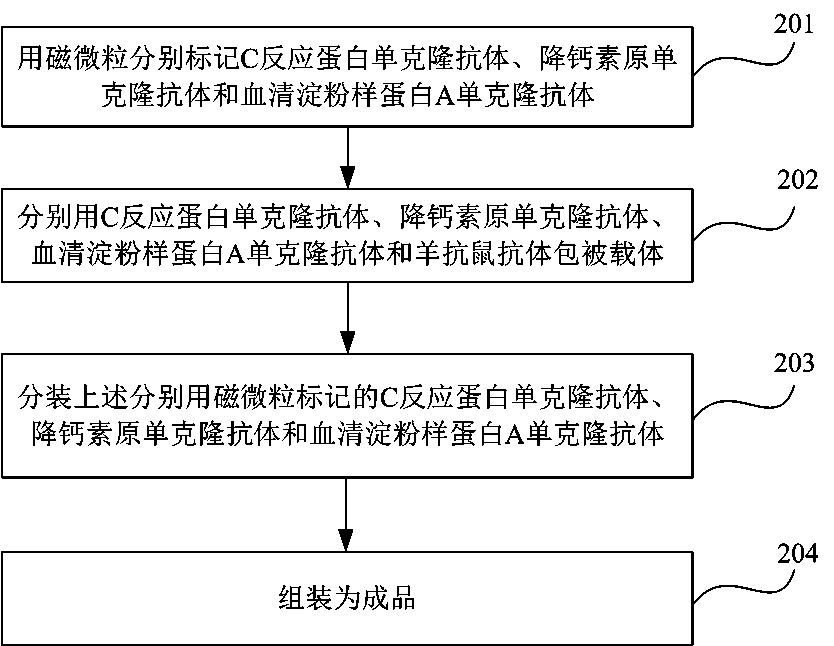
[0047] Embodiment 1 Preparation of coating film:
[0048] Coating buffer preparation: 0.05M pH 7.4 phosphate buffer (PB) was used as the coating buffer, which was sterilized by 0.22 μm microporous membrane and stored at 4°C for later use.
[0049] Preparation of blocking solution: 0.05M phosphate buffered saline (PBS) containing 1.0% bovine serum albumin (BSA) in mass ratio, pH 7.4, filtered through a 0.22 μm microporous membrane and stored at 4°C for later use.
[0050] Preparation of coating membrane: Dilute C-reactive protein / procalcitonin / serum amyloid A antibody to 0.5mg / ml with 0.05M phosphate buffer (PB), pH7.4 coating buffer, goat anti-mouse The secondary antibody was diluted to 1 mg / ml, and was uniformly spray-printed at 0.5 cm intervals on NC membranes with a width of 3.5 cm using a quantitative spraying device at 1 μl / cm. 0.05M PBS, pH 7.4) for 10 minutes, dried at 25-35°C for 8 hours, added desiccant and sealed for later use.
Embodiment 2
[0051] Example 2 Preparation of magnetic particle-labeled antibody:
[0052] Preparation of citric acid buffer: Weigh 14.7g of sodium citrate and 10.5g of citric acid and dissolve in purified water to prepare 1L of citric acid buffer with a pH value of 4.6 and a concentration of 0.05M, and add Tween-20 with a volume ratio of 0.05%. Filter and sterilize with a 0.22 μm microporous membrane and store at 4°C for later use.
[0053] Preparation of boric acid storage buffer: Weigh 0.62g of boric acid and 3.81g of borax and dissolve in purified water to prepare 1L of boric acid buffer solution with a pH of 9.0 and a final concentration of 0.05M, and add povidone (PVP) with a mass ratio of 1%. 0.5% Casein, 0.5% Tween-20 by volume, 5% sucrose by mass, sterilized by 0.22 μm microporous membrane and stored at 4°C for later use.
[0054] Preparation of magnetic particles:
[0055] 1. Preparation of magnetic particles containing CRP antibody:
[0056] (1) Wash the magnetic particles wit...
Embodiment 3
[0071] Example 3 Assembly of test strips and finished products
[0072] All of the following operating environment requirements are: humidity less than 20%, temperature 18-26 ℃.
[0073] Assembling the test board: use the BioDot LM5000 assembly machine to assemble the 3.5cm coating film on the transparent plastic base plate with a width of 9.8cm according to the requirements, cover the upper transparent plastic cover, and assemble the test board.
[0074] Cutting of test strips: Use a BioDot CM4000 strip cutter to cut the assembled test strips into 0.5cm wide test strips, then pack them into 20 strips / tube, and store them at room temperature.
[0075] D. Aliquoting of magnetic particle-labeled antibody
[0076] The prepared magnetic particles containing the three antibodies were divided into 25mL / bottles and stored at 2-8°C.
[0077] E. Finished product packaging
[0078] A tube filled with 20 test strips and a glass bottle filled with 25mL mixed magnetic particle-labeled a...
PUM
| Property | Measurement | Unit |
|---|---|---|
| Particle size | aaaaa | aaaaa |
| Sensitivity | aaaaa | aaaaa |
Abstract
Description
Claims
Application Information
 - R&D
- Intellectual Property
- Life Sciences
- Materials
- Tech Scout
- Unparalleled Data Quality
- Higher Quality Content
- 60% Fewer Hallucinations
Browse by: Latest US Patents, China's latest patents, Technical Efficacy Thesaurus, Application Domain, Technology Topic, Popular Technical Reports.
© 2025 PatSnap. All rights reserved.Legal|Privacy policy|Modern Slavery Act Transparency Statement|Sitemap|About US| Contact US: help@patsnap.com


