Thyroid tumor pathological tissue section image classification method and device

A technology for thyroid tumors and tissue slicing, applied in image analysis, image enhancement, image data processing, etc., can solve problems such as time-consuming and labor-intensive, and not clinically practical

Inactive Publication Date: 2018-10-30
FUDAN UNIV SHANGHAI CANCER CENT +1
View PDF3 Cites 45 Cited by
  • Summary
  • Abstract
  • Description
  • Claims
  • Application Information

AI Technical Summary

Problems solved by technology

This method is time-consuming and labor-intensive, and is affected by human factors such as physician experience, and even the physician who took the screenshot has alr

Method used


Image

  • Thyroid tumor pathological tissue section image classification method and device
  • Thyroid tumor pathological tissue section image classification method and device
  • Thyroid tumor pathological tissue section image classification method and device

Examples


Embodiment 1

[0068] Example 1 Obtain and classify the original image set of thyroid tumor pathological tissue slices

[0069] The pathological section data set used in the present invention comes from the actual application site. The data set comes from the Fudan University Affiliated Tumor Hospital. It is obtained from pathological section specimens prepared by resection of thyroid tumors. The hospital regards such pathological tissue sections as being It is an important diagnostic basis for diagnosing cancer, so it is of great significance to use this type of data set to build an automated diagnosis system. After we obtain the slice data set, we need to classify it first. There are 7 categories: normal, adenoma, thyroid nodule, papillary carcinoma, follicular carcinoma, medullary carcinoma and undifferentiated carcinoma. Labeled as 0-6, this is an encoding process that converts abstract information into digital information. The normal data set has 93 original pathological slice images, the...

Embodiment 2

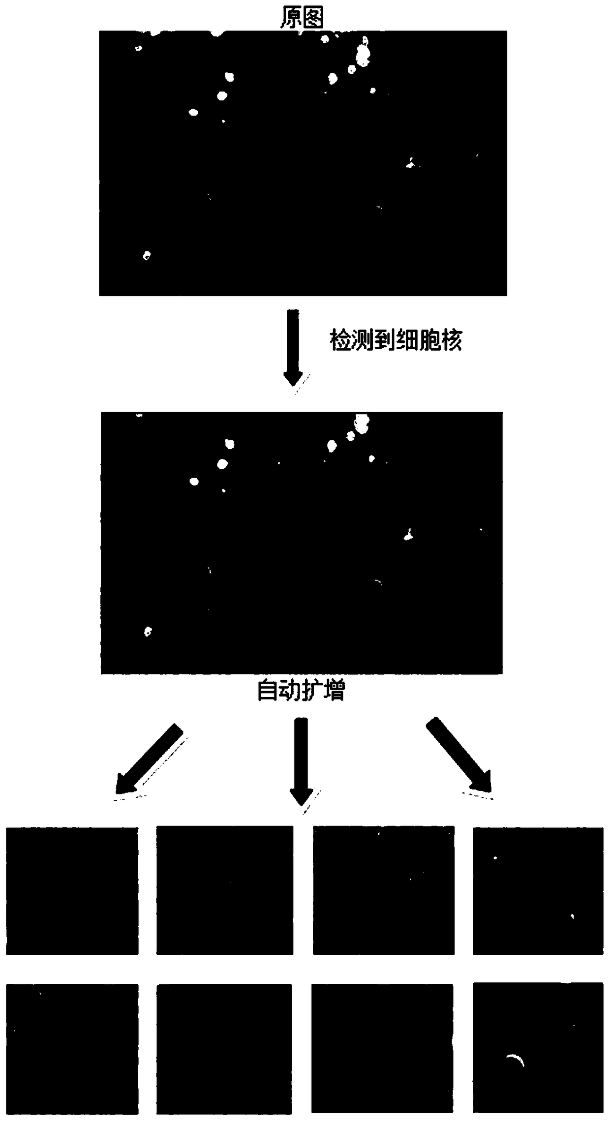
[0070] Example 2 Fully automated interception of sub-image set data amplification

[0071] Deep learning training requires a large amount of training data support to achieve satisfactory classification results. In the medical field, due to privacy protection, it is often difficult to collect a large number of data sets to meet the training requirements. Due to the limited number of data sets obtained, there are only 632, but the system needs to classify 7 categories, so we need to expand the data set. The previous amplification method was mainly used by professional doctors to find the denser tumor cells in the pathological section to intercept. According to this method, one pathological section can amplify several pictures containing certain tumor cells. The amplification method is very time-consuming and cumbersome to operate, and professional doctors often do not have enough time. Therefore, this embodiment adopts the fully automatic interception amplification method, and the ...

Embodiment 3

[0086] Example 3 Using flip and / or rotation to perform a second data augmentation on the training set

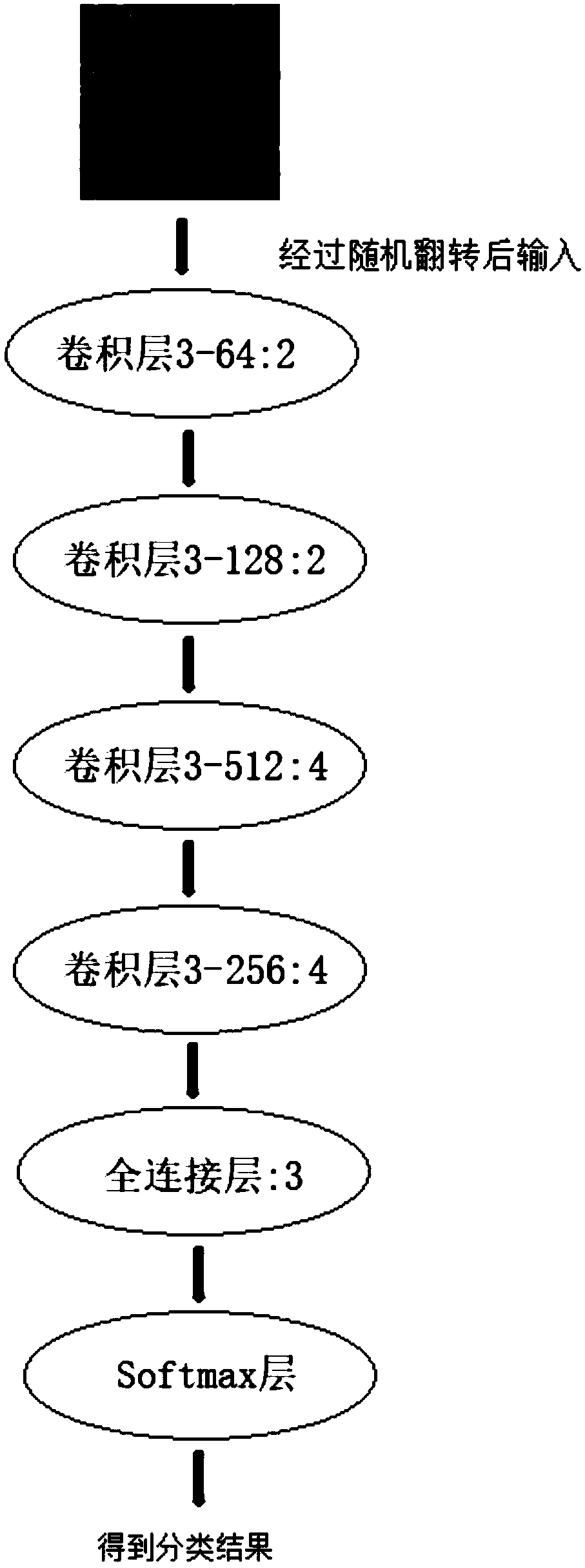
[0087] The second data augmentation of the training set is based on the horizontal flip and / or rotation of the sub-images. The horizontal flip can expand the data set to twice the original size, and rotate 0 degrees, 90 degrees, 180, and 270 degrees. The original training set is expanded four times, so the original data set can be expanded eight times after horizontal flipping and rotation.

[0088] The second amplification of the training set is to use the online data amplification method. Compared with the traditional pre-amplification method before training, this online amplification method has the advantage of reducing the occupation of the amplified data set. Storage space. It only needs to adopt the method of batch training. During training, each sub-image in each batch of training is randomly flipped and / or rotated. When the number of training iterations is large enough, ...


PUM


Abstract

The invention discloses a thyroid tumor pathological tissue section image classification method and a thyroid tumor pathological tissue section image classification device. The method comprises the steps of acquiring an original image set of classified thyroid tumor pathological tissue sections; automatically intercepting multiple area images including cells from each original image to serve as asub-image set; using the total or partial sub-image set as a training set; building a preliminary convolutional neural network model; training the preliminary convolutional neural network model by using the training set to acquire a mature convolutional neural network model; and classifying the classified thyroid tumor pathological tissue section images by using the mature convolutional neural network model. Cell nucleuses in the thyroid tumor pathological tissue sections are matched by using Gaussian laplace operator characteristics to find positions of cells, automatic image interception isimplemented in the area with many cells, so that the full-automatic cell image classification and cancer diagnosis are achieved, the workload of a doctor when in checking of the tissue section image can be greatly reduced, and the diagnosis accuracy is improved.

Description

Technical field [0001] The invention relates to the field of artificial intelligence and image recognition, in particular to a method and device for fully automatic thyroid tumor pathological tissue slice image classification based on deep learning. Background technique [0002] Deep learning is currently the most suitable and widely used algorithm for image recognition in the field of artificial intelligence. It mimics the working mechanism of the human brain. It uses a convolutional neural network to automatically extract features from external input data, thereby enabling the machine Understand learning data, obtain information and output. At present, artificial intelligence based on deep learning has been applied to various industries, including speech recognition, face recognition, vehicle logo recognition, handwritten Chinese character recognition, etc. [0003] In the process of medical diagnosis, pathological diagnosis of tumor specimens after surgical resection is the gol...

Claims


Application Information

Patent Timeline
IPC IPC(8): G06K9/62G06N3/04G06T7/00
CPCG06T7/0012G06T2207/30096G06N3/045G06F18/241
Inventor 向俊卢宏涛王立官青王蕴珺李端树王坚刘绮颖
Owner FUDAN UNIV SHANGHAI CANCER CENT
Why Eureka
Try Eureka