In vitro tumor cell 3D collective invasion model and construction method thereof

A tumor cell and construction method technology, applied in the field of in vitro tumor cell 3D collective invasion model and its construction, can solve problems such as in-depth research on unfavorable tumor invasion, the inability of cells to directly determine whether they are tumor cells, and the inability to observe cell invasion.

Active Publication Date: 2018-11-13
SOUTHERN MEDICAL UNIVERSITY
View PDF6 Cites 6 Cited by
  • Summary
  • Abstract
  • Description
  • Claims
  • Application Information

AI Technical Summary

Problems solved by technology

[0003] The traditional in vitro 3D growth model of a single tumor cell can visually observe the growth and proliferation of tumor cells in the gel, but cannot observe the cell invasion, and it is difficult to simulate the collective invasion of tumor cells in vivo; Tumor tissue blocks are multicellular tissue blocks. Although cell invasion and growth can be observed, there are also various cellular components (such as vascular endothelial cells, fibroblasts, muscle cells, etc.) in the tissue block besides tumor cells. It is not possible to directly determine whether they are tumor cells, and it is difficult to obtain tumor cells in the target area, especially in the absence of specific biological markers, it is difficult to distinguish Leader Cells and Follower Cells in collective invasive cells, which is not conducive to tumor invasion follow-up in-depth research

Method used


Image

  • In vitro tumor cell 3D collective invasion model and construction method thereof
  • In vitro tumor cell 3D collective invasion model and construction method thereof
  • In vitro tumor cell 3D collective invasion model and construction method thereof

Examples


Embodiment 1

[0049] Example 1 Preparation of tumor multicellular spheroids

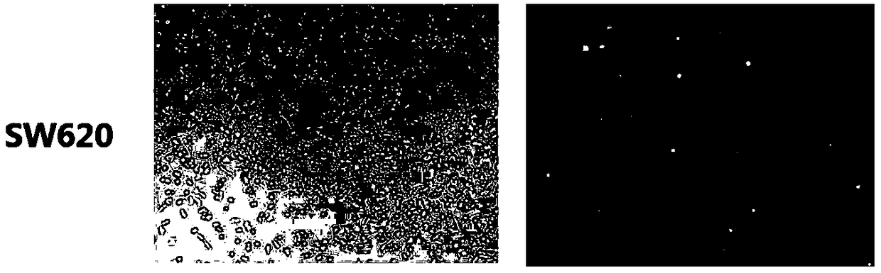
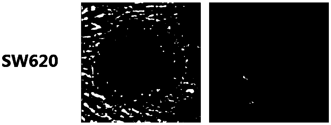
[0050] The colorectal cancer cells with good growth condition and adherent growth were washed with PBS, digested with trypsin, mixed with Gibco fetal bovine serum and Hyclone1640 medium, and resuspended in medium containing 10% fetal bovine serum. orifice plate. When plating, the cell density covers approximately 50-60% of the bottom of the well plate. Among them, two cell lines, SW620 and HT29, were selected to prepare two corresponding tumor multicellular spheroids, so as to facilitate the subsequent observation of different invasive abilities and states of different cell lines.

[0051] After the cells are in a good state of adherence and growth, phototransformation plasmids are transfected using the lipofection method, and the pDendra2-C Vector plasmids are transferred into the cells. The pDendra2-C Vector plasmids have green fluorescence; observe after 24 hours Cell fluorescence, SW620 cell line was transfe...

Embodiment 2

[0055] This example provides a method for constructing a 3D collective invasion model of tumor cells in vitro, using a laser confocal culture dish as a research device for the 3D collective invasion model of tumor cells in vitro.

[0056] In order to ensure that the growth and invasion of tumor multicellular spheroids are completely carried out in the 3D gel instead of along the glass bottom surface of the laser confocal dish, a small amount of prepared type I rat tail collagen needs to be added to the middle B area of ​​the laser confocal dish, and ensure that The distance between the tumor cell spheroid and the glass bottom of the laser confocal culture dish was greater than 100 μm on average.

[0057] Among them, the commodity information of type I rat tail collagen is: Collagen type I, rat tail.

[0058] The preparation steps of type I rat tail collagen are as follows. All glue preparation steps and reagents (type I rat tail collagen, 10×PBS, dH 2 O, 1N NaOH) should be ...

Embodiment 3

[0078] This example provides a method for constructing a 3D collective invasion model of tumor cells in vitro, using a laser confocal culture dish as a research device for the 3D collective invasion model of tumor cells in vitro. The method is basically the same as that in Example 2, the main difference is that the tumor multicellular spheroids used in Example 3 are prepared by the hanging drop culture method.

[0079] Tumor multicellular spheroids prepared by hanging drop culture figure 2 Similar, but because the number of cells in the tumor cell spheroids prepared by the hanging drop method is difficult to control, this in vitro 3D collective invasion model of tumor cells can be used for qualitative analysis, but it is difficult to achieve quantitative analysis.


PUM


Abstract

The invention provides an in vitro tumor cell 3D collective invasion model and a construction method thereof. Tumor multicellular organ groups are prepared; cells contain light conversion fluorescentprotein which is coated into type I rat-tail collagen for carrying out 3D culture and the growth state, a proliferation mode and an invasion mode of tumor cells are simulated in vitro. The model studies the collective invasion effect of tumor multicellular spherical organs and can simulate the collective invasion mode of the tumor cells in vivo; after photobleaching is carried out by using a laserconfocal microscope, Leader Cells and Follower Cells in the collective invasion cells can be distinguished under the condition of lacking specific biological markers; in addition, the tumor cells ina target region can be obtained by fluorescence flow separation for carrying out follow-up experimental study.

Description

technical field [0001] The invention relates to the field of medical biology, in particular to an in vitro tumor cell 3D collective invasion model and a construction method thereof. Background technique [0002] In the traditional observation of 3D model of tumor cells in vitro, one is to coat single tumor cells in matrigelmatrix glue or type I mouse tail collagen for culture and observation; Small pieces of tissue were coated with type I rat tail collagen for culture and observation. [0003] The traditional in vitro 3D growth model of a single tumor cell can visually observe the growth and proliferation of tumor cells in the gel, but it cannot observe the cell invasion, and it is difficult to simulate the collective invasion of tumor cells in vivo; Tumor tissue mass is a multicellular tissue mass. Although cell invasion and growth can be observed, in addition to tumor cells, the tissue mass is also mixed with a variety of cellular components (such as vascular endothelial ...

Claims


Application Information

Patent Timeline
Patent Type & Authority Applications(China)
IPC IPC(8): C12N5/10
CPCC12N5/0693C12N2533/54C12N2513/00C12N2510/00
Inventor 梁莉王斐斐朱孝辉丁彦青
Owner SOUTHERN MEDICAL UNIVERSITY
Why Eureka
Try Eureka