Eureka AIR delivers breakthrough ideas for toughest innovation challenges, trusted by R&D personnel around the world.

Detection method based on abdominal CT medical image fusion classification

A medical image and detection method technology, applied in the field of medical image processing, can solve problems such as poor noise resistance, long running time, and high time complexity, and achieve effective class label classification, reduce the risk of misjudgment, and effective feature selection.

Active Publication Date: 2018-11-27
济南市第四人民医院
View PDF3 Cites 9 Cited by
  • Summary
  • Abstract
  • Description
  • Claims
  • Application Information

AI Technical Summary

Problems solved by technology

In terms of image decomposition and reconstruction methods, image fusion methods based on the frequency domain use Fourier transform and inverse Fourier transform to decompose and reconstruct image signals at different scales, but this type of method has high time complexity and runs The characteristics of long time, which contradicts the high real-time medical aided diagnosis, and has very high requirements on the hardware and software facilities of the experimental platform.
Later, researchers proposed to use spatial domain filters to process images to perform multi-scale decomposition and reconstruction of input images. Although this type of method can quickly perform image decomposition and reconstruction, the image fusion method based on spatial domain has poor noise immunity.
However, the existing methods use the same feature for fusion of medical images of different modalities.

Method used


Image

  • Detection method based on abdominal CT medical image fusion classification
  • Detection method based on abdominal CT medical image fusion classification
  • Detection method based on abdominal CT medical image fusion classification

Examples


Embodiment Construction

[0018] The technical solutions in the embodiments of the present invention will be described clearly and in detail below with reference to the drawings in the embodiments of the present invention. The described embodiments are only some of the embodiments of the invention.

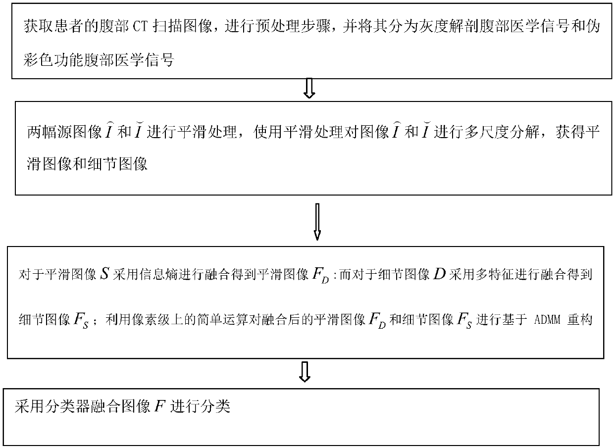
[0019] The technical scheme that the present invention solves the problems of the technologies described above is: figure 2Shown is a detection method based on abdominal CT medical image fusion classification, which is used to process the patient's abdominal CT medical image to obtain the patient's suspected lesion area, which includes the following steps: the acquisition step, acquiring the patient's abdomen CT scanning images, start the CT scanner to perform cine-mode CT scanning on the abdomen of the human body, and assign bed numbers and layer numbers to each CT image according to the order in which the human body enters the scanning area. , assign a phase number to each picture of the CT image accor...


PUM


Abstract

The present invention claims a detection method based on abdominal CT medical image fusion classification. The detection method comprises: an acquisition step of acquiring an abdominal CT scanning image of a patient; a pre-processing step of performing pre-processing to obtain a binarized grayscale image; a morphological corrosion and dilation step of performing morphological corrosion calculationto obtain a corroded image, and performing dilation operation on the binarized grayscale image to obtain a dilated image; an operation step of performing opening operation on the corroded image to obtain an open operation map and performing closed operation on the dilated image to obtain a closed operation map; a Fourier transform step of obtaining a Fourier transform value; an abdominal medicalimage fusion classification step for performing fusion classification distinguishing on the abdominal CT scanning image; and a lesion detection step of performing determination according to the Fourier transform value and the fusion classification distinguishing result to obtain a suspected lesion area of the patient. The invention can lower the complexity and improve the accuracy of recognition and diagnosis of abdominal lesions.

Description

technical field [0001] The invention belongs to the technical field of medical image processing, in particular to a detection method based on abdominal CT medical image fusion classification. Background technique [0002] The existing abdominal medical image fusion technology is mainly for two different modal medical images. According to the different medical image modalities, the medical image fusion system can be divided into three types: anatomical medical image and anatomical medical image fusion, anatomical medical image and anatomical medical image fusion. Functional medical image fusion and functional medical image and functional medical image fusion. The MRI-PET and MRI-SPECT medical image fusion system belongs to the fusion of anatomical medical images and functional medical images. The input images of this system are grayscale and pseudo-color. The MRI-PET combination kit launched by Philips combines a commercial MRI imaging scanner with a PET with special shieldi...

Claims


Application Information

Patent Timeline
IPC IPC(8): G06T7/00
CPCG06T7/0012G06T2207/10081G06T2207/20056G06T2207/20221
Inventor 成英
Owner 济南市第四人民医院
Why Eureka
Eureka Blog
Learn More