Method for analyzing load rate of graphene oxide for nucleic acid

An analysis method, graphene technology, applied to the analysis of materials, material excitation analysis, material analysis through optical means, etc., to achieve the effect of simple calculation and convenient operation

Inactive Publication Date: 2018-12-11
THE SECOND HOSPITAL AFFILIATED TO WENZHOU MEDICAL COLLEGE
View PDF5 Cites 0 Cited by
  • Summary
  • Abstract
  • Description
  • Claims
  • Application Information

AI Technical Summary

Problems solved by technology

Unfortunately, so far there are no literature reports on the analysis...

Method used


Image

  • Method for analyzing load rate of graphene oxide for nucleic acid
  • Method for analyzing load rate of graphene oxide for nucleic acid
  • Method for analyzing load rate of graphene oxide for nucleic acid

Examples


Embodiment 1

[0024] Example 1: An example of analyzing the loading rate of GO to a kind of green fluorescent double-stranded RNA.

[0025] The sequence of the double-stranded RNA is shown in the appendix figure 2 As shown, the molar mass M is 12112 g / mol, the fluorescent modification group is FAM, the emission wavelength is 480 nm, and the excitation wavelength is 520 nm.

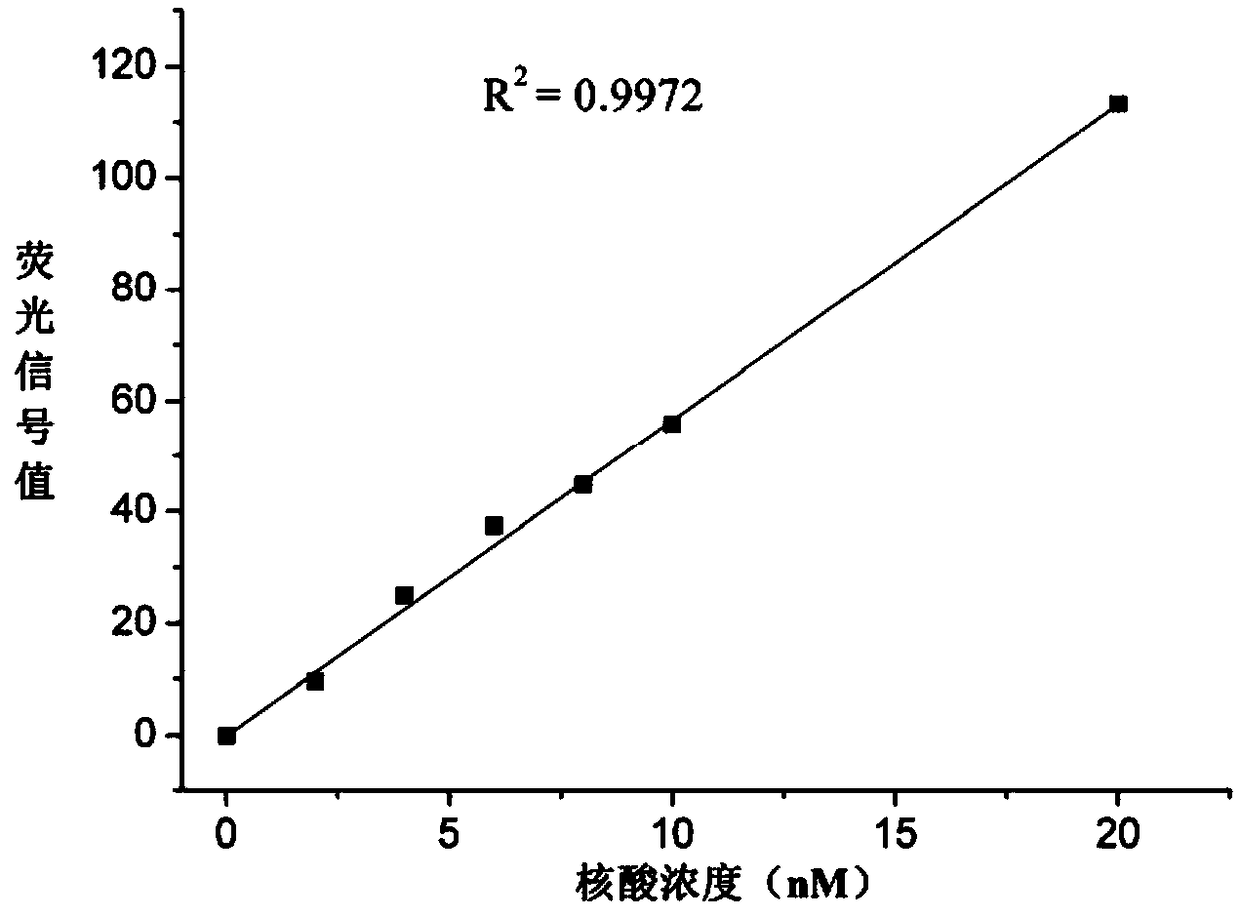
[0026] The specific implementation process is as follows: prepare DEPC solutions with concentrations of 0, 2, 4, 6, 8, 10, and 20 nM of the nucleic acid respectively, and use a fluorescence spectrophotometer (Shanghai Prism Technology Co., Ltd., model F96PRO, emission wavelength 480 nm, Excitation wavelength 520nm) for detection, obtain the standard fluorescence signal values ​​of the nucleic acid solution to be tested at each concentration, and draw a standard curve ( figure 1 ). Bring the volume to 1.6 mL with an initial concentration of n 1 The DEPC solution of the nucleic acid (treated with high-temperature and...

Embodiment 2

[0030] Example 2: An example of analyzing the loading rate of GO on a red fluorescent double-stranded RNA.

[0031] The sequence of the double-stranded RNA is attached Figure 4 , the molar mass M is 12112 g / mol, the fluorescent modification group is CY3, the emission wavelength is 550 nm, and the excitation wavelength is 570 nm.

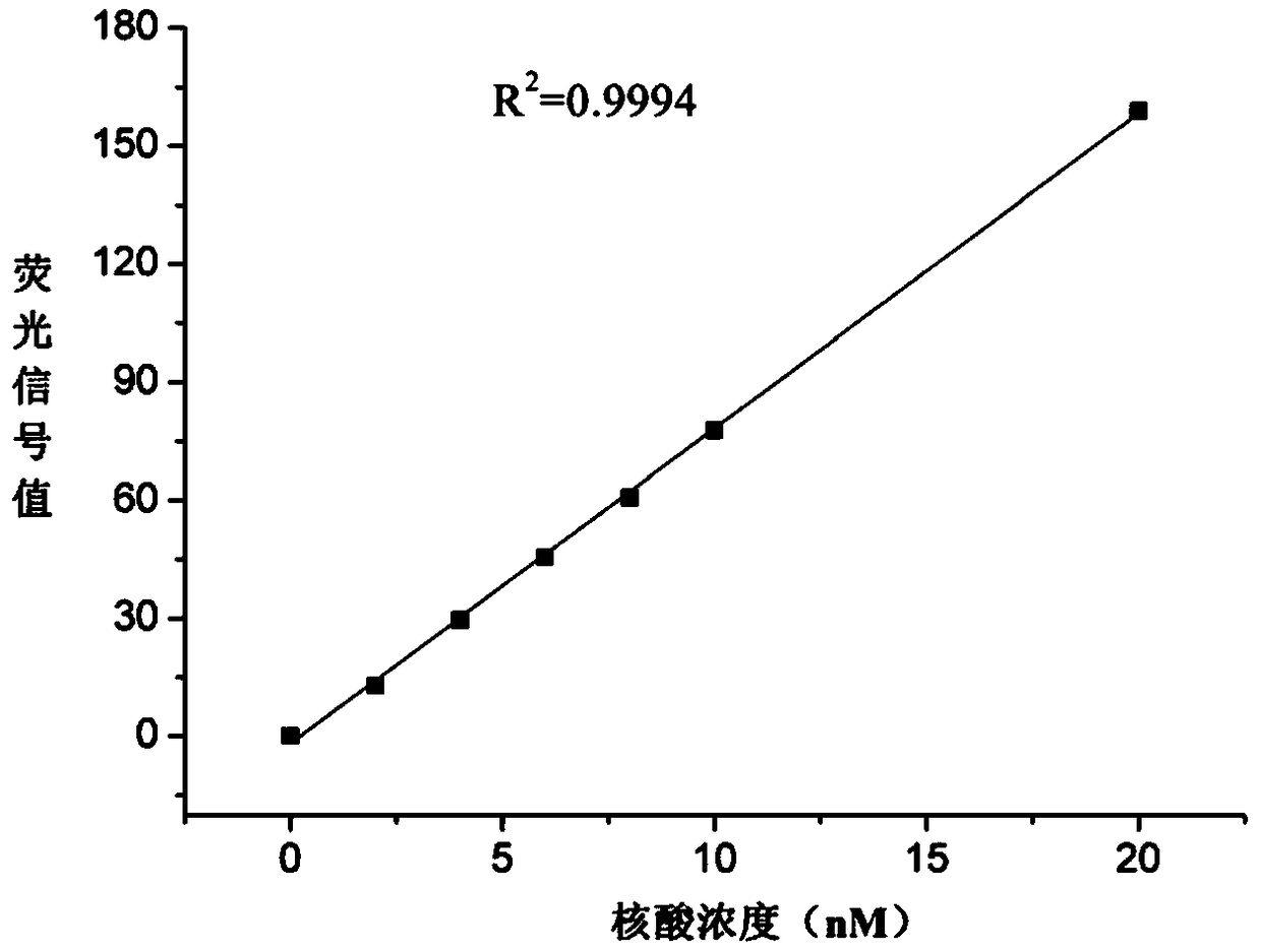
[0032] The specific implementation process is as follows: prepare DEPC solutions with concentrations of 0, 2, 4, 6, 8, 10, and 20 nM of the nucleic acid respectively, and use a fluorescence spectrophotometer (Shanghai Prism Technology Co., Ltd., model F96PRO, emission wavelength 550 nm, Excitation wavelength 570nm) for detection, obtain the standard fluorescence signal values ​​of the nucleic acid solution to be tested at each concentration, and draw a standard curve ( figure 2 ). Bring the volume to 1.6 mL with an initial concentration of n 1 The DEPC solution of the nucleic acid and the DEPC solution of GO (20 μg / mL) were mixed in equal volume...

Embodiment 3

[0036] Example 3: An example of analyzing the loading rate of GO to a kind of green fluorescent single-stranded DNA.

[0037] The single-stranded DNA sequence is attached Figure 6 , the molar mass M is 23016 g / mol, the fluorescent modification group is FAM (green fluorescence), the emission wavelength is 480 nm, and the excitation wavelength is 520 nm.

[0038] The specific implementation process is as follows: prepare TE solutions with concentrations of 0, 2, 4, 6, 8, 10, and 20 nM of the nucleic acid respectively, and use a fluorescence spectrophotometer (Shanghai Prism Technology Co., Ltd., model F96PRO, emission wavelength 480 nm, Excitation wavelength 520 nm) for detection, obtain the standard fluorescence signal values ​​of the nucleic acid solution to be tested at each concentration, and draw a standard curve ( image 3 ). Bring the volume to 1.6 mL with an initial concentration of n 1 The TE solution of the nucleic acid and the TE solution of GO (20 μg / mL) were mix...


PUM


Abstract

The invention discloses a method for analyzing a load rate of graphene oxide for nucleic acid. The method comprises the following steps: (1) drawing a fluorescence signal standard curve of nucleic acid, (2) performing quenching fluorescence treatment on graphene oxide, and (3) calculating the load rate of graphene oxide for nucleic acid, wherein the load rate L is a ratio of loaded nucleic acid mass to graphene oxide mass; the computational formula is as follows: L=(C1-C2)V/C GOV*%=(C1-C2)/CGO*%=(n1-n2)M/CGO*%; C1 and C2 are respectively mass concentrations of nucleic acid in the solution before and after load; CGO is the mass concentration of GO in the solution; V is the volume of solution; n1 and n2 are respectively molar concentrations of nucleic acid in the solution before and after load; M is the molar mass of nucleic acid. The invention has the advantages that the method is capable of quickly realizing the quantitative analysis and calculation for nucleic acid molecules loaded bygraphene oxide on the basis of the fluorescence quenching characteristic of graphene oxide.

Description

technical field [0001] The invention belongs to the field of biomacromolecule analysis, and specifically refers to a method for quantitative analysis and calculation of nucleic acid molecules carried by graphene oxide. Background technique [0002] Graphene is made of carbon atoms with sp 2 A two-dimensional periodic honeycomb lattice structure formed by hybrid orbitals. Graphene is the basic unit that constitutes other graphite materials, and can construct zero-dimensional fullerenes, one-dimensional carbon nanotubes and three-dimensional bulk graphite. The basic structural unit of graphene is the most stable benzene six-membered ring in organic materials, so it is currently the most ideal two-dimensional nanomaterial. Graphene materials have many excellent properties, such as good electrical and thermal conductivity, ultra-high mechanical strength, and unique electrical properties. Graphene and its derivatives have been widely used in many fields including materials sci...

Claims


Application Information

Patent Timeline
IPC IPC(8): G01N21/64
CPCG01N21/6428G01N2021/6432
Inventor 李磊褚茂平陈永辉刘畅林振坤王洁王瑶尧曾静静金其可
Owner THE SECOND HOSPITAL AFFILIATED TO WENZHOU MEDICAL COLLEGE
Why Eureka
Try Eureka