Fluorescent carbon dot for nuclear staining, and application and application method of same to nuclear imaging

A fluorescent carbon dot and fluorescent imaging technology, applied in the field of nanomaterials and biological imaging, can solve the problems of long incubation time and difficult cell nucleus imaging, and achieve the effects of short incubation time, cheap devices and reagents, and low incubation concentration

Active Publication Date: 2018-12-18
ZHENGZHOU UNIV
View PDF3 Cites 6 Cited by
  • Summary
  • Abstract
  • Description
  • Claims
  • Application Information

AI Technical Summary

Problems solved by technology

When the carbon dots invented at present are used for cell imaging, due to the size of the material, surface charge and other properties, it is difficult to apply to the imaging of the nucleus. Most of the invented carbon dots can only enter the cytoplasm, mitochondria and other parts of the cell, and require longer incubation time

Method used


Image

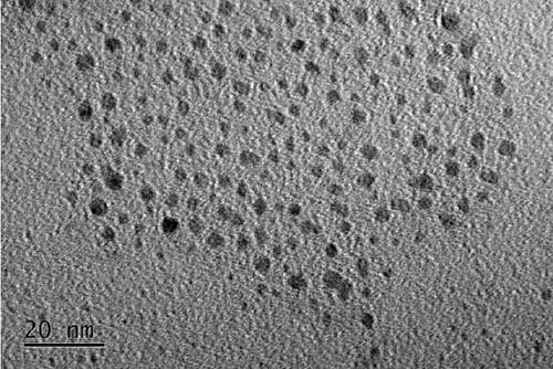
  • Fluorescent carbon dot for nuclear staining, and application and application method of same to nuclear imaging
  • Fluorescent carbon dot for nuclear staining, and application and application method of same to nuclear imaging
  • Fluorescent carbon dot for nuclear staining, and application and application method of same to nuclear imaging

Examples


Embodiment 1

[0029] A fluorescent carbon dot for nuclear staining, the carbon dot is prepared by the following steps:

[0030] 1) Dissolve 5 mg of folic acid and 13 mg of m-phenylenediamine in 5000 mg of deionized water, place in a hydrothermal reactor and heat to 200°C for 12 hours; in other examples, the mass ratio of folic acid and m-phenylenediamine is 1 : (2-20) can be used, the amount of deionized water can be enough to dissolve the raw materials;

[0031] 2) After the reaction, centrifuge (remove large particle aggregates) and take the upper layer solution through silica gel column chromatography, collect the yellow-green fluorescent part, spin evaporate (remove organic solvent), transfer to distilled water, freeze-dry to obtain fluorescent carbon dots; The mobile phase used in silica gel column chromatography is a mixture of methanol and ethyl acetate at a volume ratio of 1:1; the specific operation of lyophilization is: first freeze at -16°C for 30h, and then freeze at -60°C for 4...


PUM

PropertyMeasurementUnit
Particle sizeaaaaaaaaaa
The average particle sizeaaaaaaaaaa

Abstract

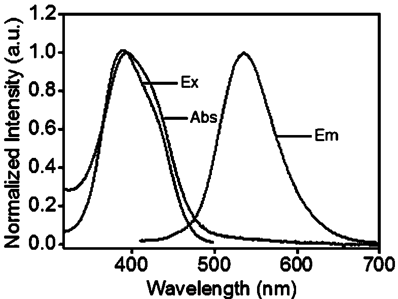
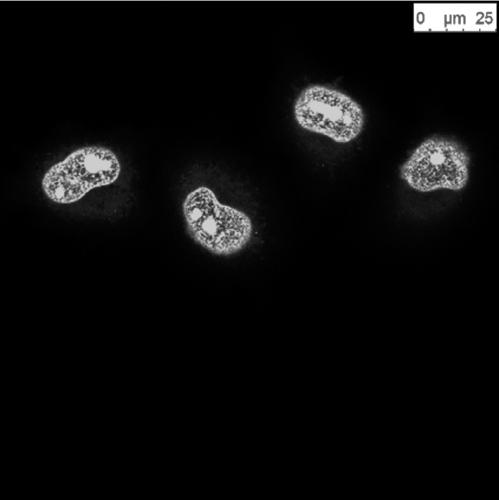
The invention provides a fluorescent carbon dot for nuclear staining. The fluorescent carbon dot is prepared from the following steps: 1) dissolving folic acid and m-phenylenediamine in water, and carrying out heating for a reaction; and 2) after the reaction, performing centrifuging and silica gel column chromatography, collecting a yellow-green fluorescent part, transferring the yellow-green fluorescent part into water after rotary evaporation and then carrying out lyophilization. The carbon dot prepared in the invention emits green light under ultraviolet excitation, has large Stokes shift,is consistent with the existing commercial 405 laser at the optimal excitation position, and can be well applied to a laser confocal microscope.

Description

technical field [0001] The invention belongs to the technical field of nanomaterials and biological imaging, and in particular relates to a fluorescent carbon dot for cell nucleus staining and its application and method in cell nucleus fluorescence imaging. Background technique [0002] Cells are important carriers in life activities, and the study of cells and foreign substances can help us understand the transportation, metabolism and toxicity of cells. Among them, the nucleus is particularly important for many processes of cells, such as metabolism, generation and inheritance. However, relative to its vital importance, the nucleus is relatively poorly studied. The staining of the nucleus is the first step, he can show the shape of the nucleus, and then the transport of the nucleus and the transport of reagents to the nucleus can be studied. With the rapid development of fluorescence microscopy, fluorescent staining of cell nuclei has become extremely common due to its v...

Claims


Application Information

Patent Timeline
IPC IPC(8): C09K11/65C01B32/15B82Y20/00B82Y40/00G01N21/64
CPCB82Y20/00B82Y40/00C09K11/65C01B32/15G01N21/6486
Inventor 李朝辉刘海芳孙远强杨杰屈凌波
Owner ZHENGZHOU UNIV
Why Eureka
Try Eureka