Image control system, method, terminal, storage medium for tumor interventional device

An image control and tumor technology, applied in the medical field, can solve the problems of inability to quickly and accurately locate images, unfavorable tumor treatment, unclear images, etc.

Pending Publication Date: 2018-12-18
刘杭
View PDF0 Cites 5 Cited by
  • Summary
  • Abstract
  • Description
  • Claims
  • Application Information

AI Technical Summary

Problems solved by technology

[0005] The images collected for tumors cannot be used for deeper and more comprehensive observations, which is not conducive to the treatment of tumors
[0006] Traditional ultrasound system cannot satisfy the acquisition of shear wave
[0007] The

Method used


Image

  • Image control system, method, terminal, storage medium for tumor interventional device
  • Image control system, method, terminal, storage medium for tumor interventional device
  • Image control system, method, terminal, storage medium for tumor interventional device

Examples


Embodiment Construction

[0111] In order to further understand the content, features and effects of the present invention, the following examples are given, and detailed descriptions are given below with reference to the accompanying drawings.

[0112] The structure of the present invention will be described in detail below in conjunction with the accompanying drawings.

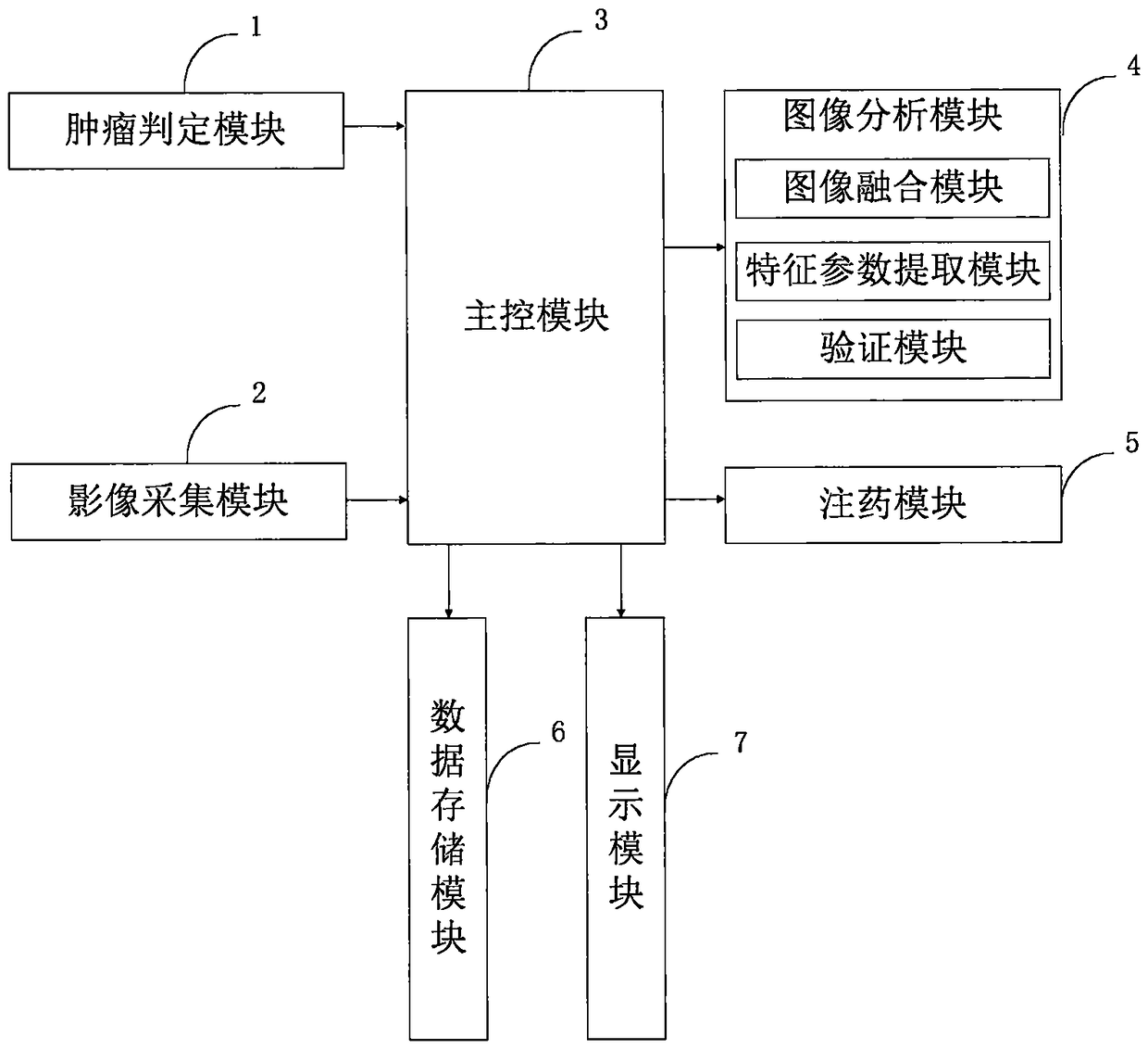
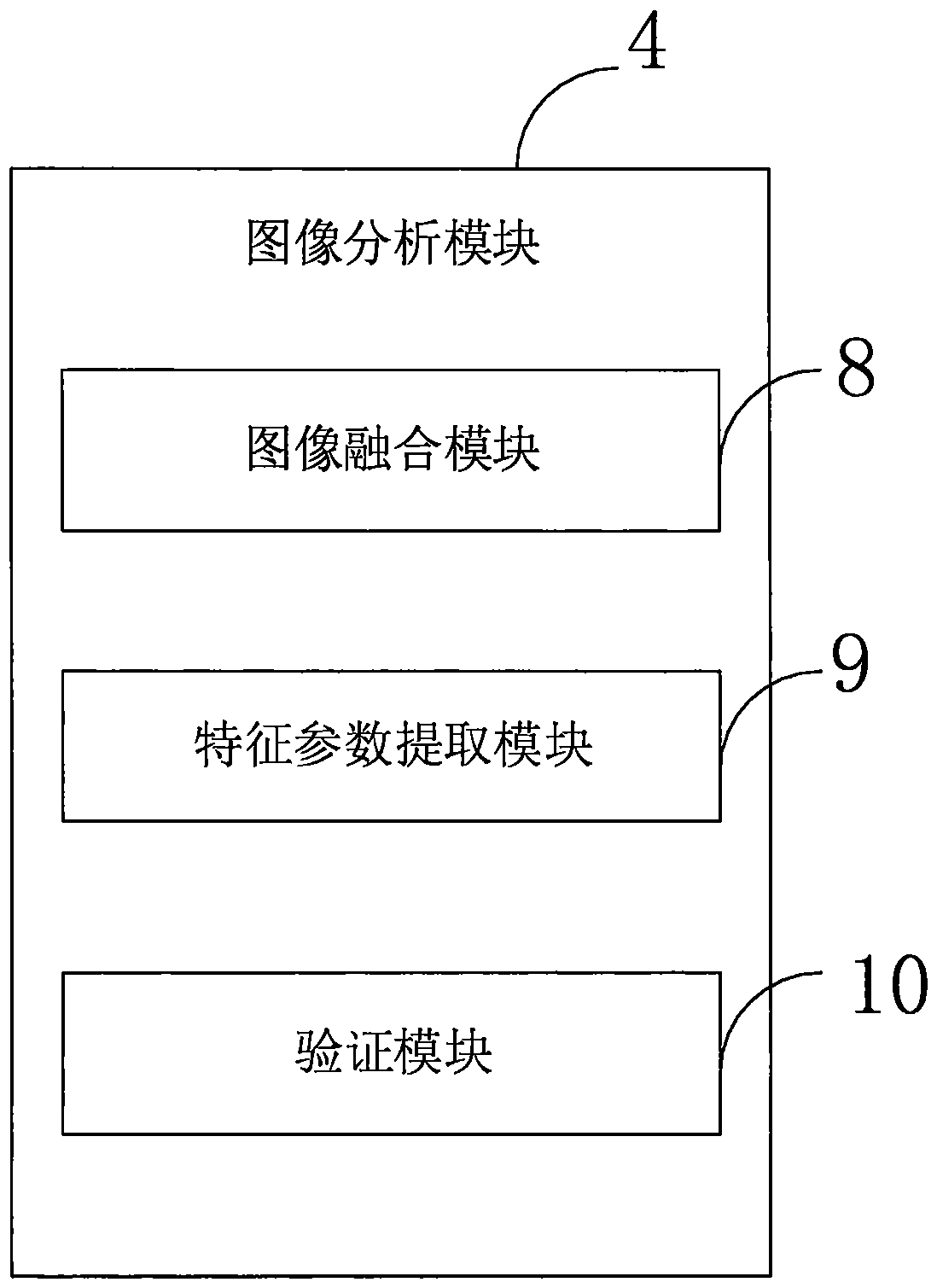
[0113] like figure 1 As shown, the image control system of the tumor interventional device provided by the embodiment of the present invention includes: a tumor determination module 1, an image acquisition module 2, a main control module 3, an image analysis module 4, a drug injection module 5, a data storage module 6, and a display module 7.

[0114] Tumor judging module 1, connected with main control module 3, for judging tumor image type;

[0115] The image acquisition module 2 is connected with the main control module 3, and is used to obtain the elastic image of the detected part through ultrasonic shear wave elastography;

...


PUM


Abstract

The invention belongs to the field of medical technology, and discloses an image control system of a tumor intervention device, a method, a terminal, a storage medium, and an image control system of the tumor intervention device, comprising a tumor judgment module, an image acquisition module, a main control module, an image analysis module, a drug injection module, a data storage module and a display module, wherein the image control system comprises a tumor judgment module, an image acquisition module, a main control module, an image analysis module, a drug injection module, a data storage module and a display module. The invention can judge the type of tumor image more quickly, simply and accurately through the tumor judging module, so that an appropriate treatment method can be selected for the patient. The invention is helpful for improving the survival rate of the patient; at the same time, the image analysis module combines the ultrasonic imaging hardness information with the blood oxygen information provided by the photoacoustic imaging to analyze the tumor image. The imaging information and the ultrasonic technology can form an important complement to the tissue shape andstructure, hardness information, and the combination of the two can realize the comprehensive observation of tumor structure and function.

Description

technical field [0001] The invention belongs to the field of medical technology, and in particular relates to an image control system, method, terminal and storage medium of a tumor interventional device. Background technique [0002] At present, the existing technologies commonly used in the industry are as follows: [0003] Tumor refers to the neogrowth formed by the proliferation of local tissue cells in the body under the action of various tumorigenic factors, because this neogrowth is mostly in the form of space-occupying blocky protrusions, also known as neoplasm (neoplasm) . Interventional radiology, also known as interventional therapy, is a new discipline that has developed rapidly in recent years and integrates imaging diagnosis and clinical treatment. Under the guidance and monitoring of imaging equipment such as digital subtraction angiography, CT, ultrasound, and magnetic resonance, it uses puncture needles, catheters, and other interventional equipment to int...

Claims


Application Information

Patent Timeline
IPC IPC(8): G06K9/62
CPCG06V2201/03G06F18/25G06F18/259G06F18/214
Inventor 刘杭
Owner 刘杭
Why Eureka
Try Eureka