A fundus light field imaging method and device

A technology of light field imaging and light field image, applied in the field of image processing, can solve the problems such as inability to apply fundus imaging system, inaccurate focus, edge imaging distortion, etc., to achieve convenient shooting and post-diagnosis analysis, simple transformation method, and easy promotion Effect

Active Publication Date: 2021-05-04
CHINA MOBILE COMM LTD RES INST +1
View PDF6 Cites 0 Cited by
  • Summary
  • Abstract
  • Description
  • Claims
  • Application Information

AI Technical Summary

Problems solved by technology

[0010] Embodiments of the present invention provide a fundus light field imaging method and device to solve the problem of inaccurate focus in the existing fundus optical imaging system, and the shooting is easily affected by the patient's eye movement, concave shape of the fundus, and edge imaging distortion. The problem that the existing light field imaging method cannot be applied to the fundus imaging system

Method used


Image

  • A fundus light field imaging method and device
  • A fundus light field imaging method and device
  • A fundus light field imaging method and device

Examples


Embodiment Construction

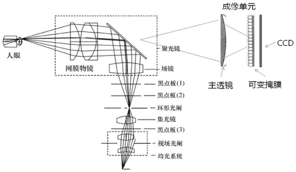
[0038] In order to solve the problem that the existing fundus optical imaging system has inaccurate focus, and the shooting is easily affected by the patient's eye movement, concave shape of the fundus and edge imaging distortion, but the existing light field imaging method cannot be applied to the fundus imaging system. In the embodiment of the present invention, a fundus light field imaging method is redesigned. The method is as follows: firstly, based on the target eye, the aperture mode of the programmable liquid crystal dynamic mask is determined, wherein the programmable liquid crystal dynamic mask includes several liquid crystal unit, and then, based on the lesion degree of the target eye, determine the spatial distribution of several liquid crystal units included in the programmable liquid crystal dynamic mask in the selected aperture mode, and based on the spatial distribution of several liquid crystal units, determine when the lighting conditions are met , start shoot...


PUM


Abstract

The present invention relates to the technical field of image processing, and in particular to a fundus light field imaging method and device, which are used to transform the existing fundus optical imaging system so that light field imaging can be applied to fundus imaging. part, determine the aperture mode of the programmable liquid crystal dynamic mask, and then determine the spatial distribution of each liquid crystal unit in the selected aperture mode based on the lesion degree of the target eye, and then determine that when the illumination is sufficient, start shooting and obtain the initial The fundus light field image, and the initial fundus light field image are processed by the preset refocusing rules to obtain the target fundus light field image. In this way, by adding a programmable liquid crystal dynamic mask, the fundus light field imaging can be realized and the fundus image can be taken When shooting, not only does not need to focus, but also does not need to be affected by the subject's eye movement, which is convenient for shooting and post-diagnosis analysis, thereby improving the user experience. Moreover, the transformation method is relatively simple and easy to promote.

Description

technical field [0001] The invention relates to the technical field of image processing, in particular to a fundus light field imaging method and device. Background technique [0002] In clinical medicine, ophthalmologists use ophthalmoscopes, fundus cameras and other instruments to observe the condition of the patient's fundus to help diagnose the disease. Since the eye itself is a complex optical system, the fundus camera needs to pass through the optical part of the eye to capture a clear fundus The imaging system of a complete fundus camera usually consists of three parts: an illumination subsystem, an observation subsystem, and a camera subsystem. [0003] At present, the fundus camera is generally an optical imaging fundus camera. Because traditional optical imaging can only record the light from the observation target, pass through the imaging aperture, and radiate to the two-dimensional imaging plane, and lose other dimensional information. The particularity of the ...

Claims


Application Information

Patent Timeline
Patent Type & Authority Patents(China)
IPC IPC(8): A61B3/14A61B3/15A61B3/12
CPCA61B3/0008A61B3/12A61B3/14A61B3/152
Inventor 信伦许利群
Owner CHINA MOBILE COMM LTD RES INST
Why Eureka
Try Eureka