

Analysis method based on multimodal image data and application thereof
A technology of image data and analysis methods, applied in the field of medical image processing and its application, can solve the problems of single target, ignoring the importance of individualized intervention treatment plan, etc.
- Summary
- Abstract
- Description
- Claims
- Application Information
AI Technical Summary
Problems solved by technology
Method used
Image

Examples
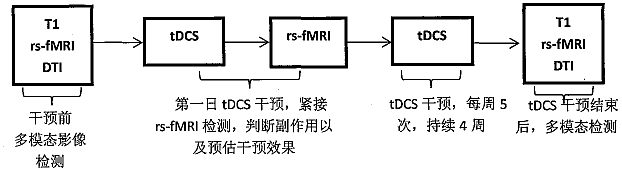
Embodiment 1
[0035] Embodiment 1 clinical trial research
[0036] 1) Determine the reference parameters of multimodal image monitoring:
[0037] High-resolution T1-weighted structural imaging (T1-weighted): echo time (TE) 2.52 milliseconds; repetition time (TR) 1900 milliseconds; inversion time (TI) 900 milliseconds; inversion angle 9 degrees; slice 176; thickness 1 mm; resolution 256×256, voxel size 1×1×1 cubic millimeter;
[0038] Resting state functional imaging: echo time (TE) 30 milliseconds; repetition time (TR) 2000 milliseconds; inversion time (TI) 900 milliseconds; inversion angle 90 degrees; slice 32; slice thickness 3 mm; slice interval 1 mm ;Resolution 64×64, voxel size 3×3×3 mm3;
[0039] Diffusion tensor imaging: A standard 8-channel head coil was used, and DTI scanning was performed using a single-shot spin-echo planar sequence (SS-EPI). TR / TE=5000 / 60 milliseconds, layer thickness 2 mm, interval 0 mm, number of excitations 2, number of layers 60, field of view (FOV): 230×...
PUM
 Abstract
Description
Claims
Application Information