Isolation and culture method of primary cervical cancer cells
A technology of primary cells and culture methods, applied in the field of cervical cancer tumor primary cell separation and culture, can solve the problems of few types and can not fully meet the research needs, and achieves long time, shortened time period and high growth efficiency. Effect
- Summary
- Abstract
- Description
- Claims
- Application Information
AI Technical Summary
Problems solved by technology
Method used
Image
Examples
Embodiment 1
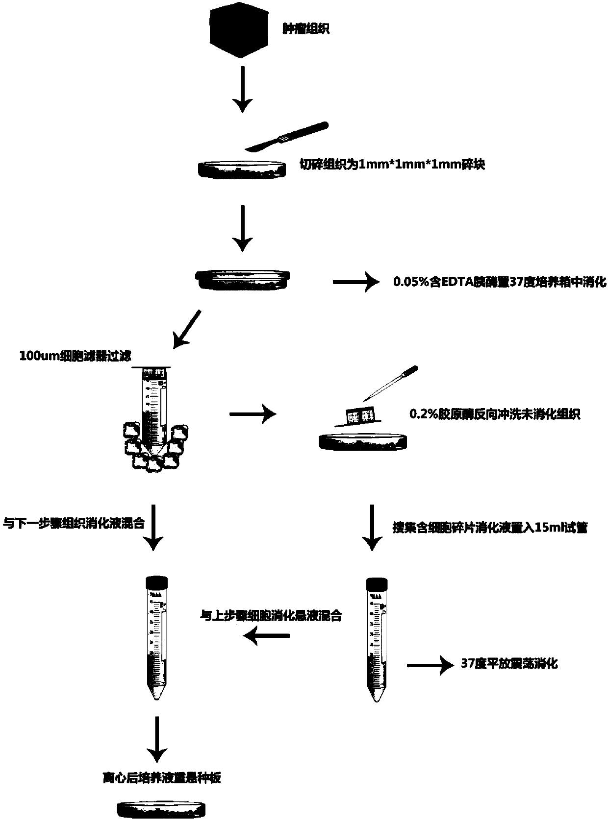
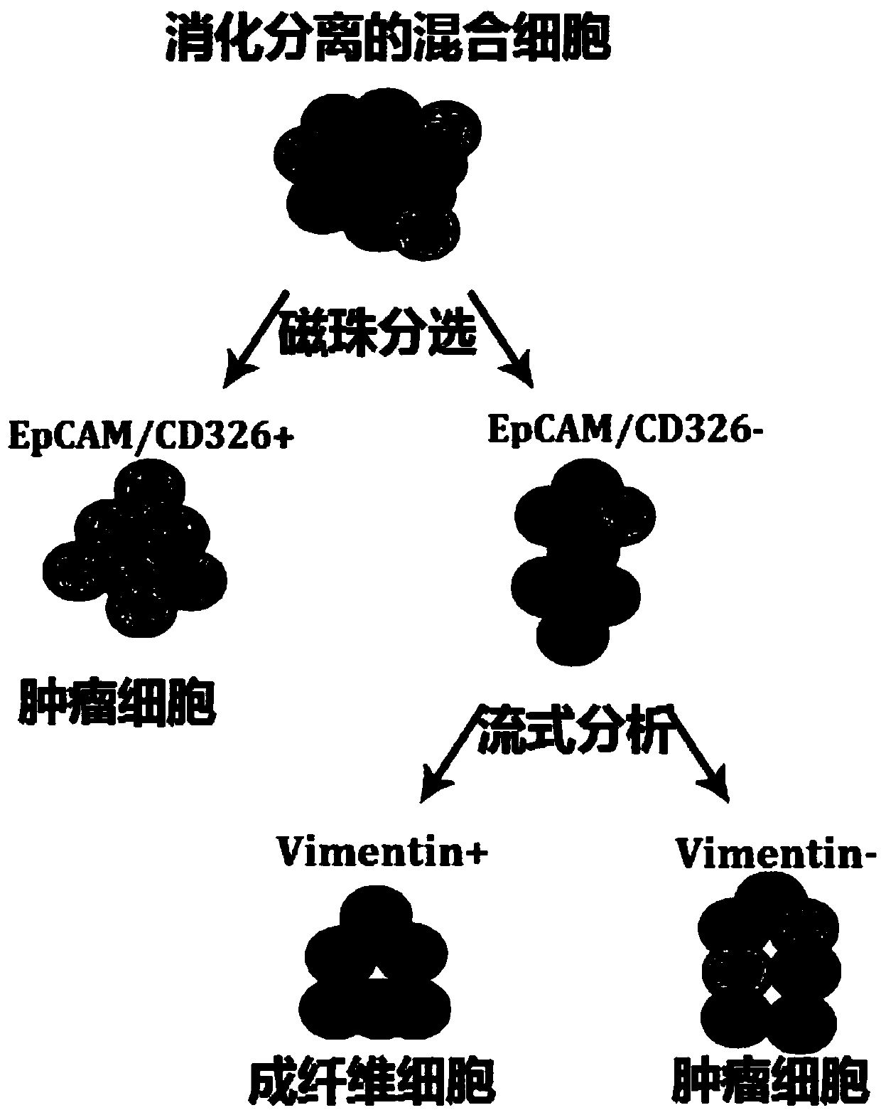
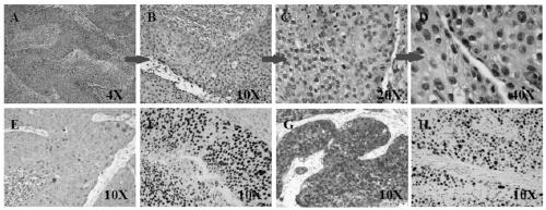
[0042] 1) Put the resected tissue block into the pre-cooled culture medium containing DMEM / F12 + 10% fetal bovine serum + 1% antibiotic, and store it at 4°C. The results of HE staining of cervical squamous cell carcinoma tissue are as follows: image 3 As shown, A, B, C and D are HE staining of cervical squamous cell carcinoma tissue, E is mouse anti-human AE1 / AE3 antibody staining (10X), I is mouse anti-human P63 antibody staining (10X), G is Mouse anti-human P16 antibody staining (10X), H is mouse anti-human Ki67 antibody staining (10X), it is clearly cervical squamous cell carcinoma, and the ratio of cervical squamous cell carcinoma tissue components is as follows Figure 4As shown, cervical cancer tissue was co-stained with mouse anti-human AE1 / AE3, rabbit anti-human Vimentin and DAPI;
[0043] 2) Wipe the blood of the tissue block with gauze in the ultra-clean bench, and rinse it with PBS+10% antibiotics, put the cleaned tissue block into a 6cm petri dish with 0.5mL DMEM-...
PUM
 Abstract
Description
Claims
Application Information
 - R&D
- Intellectual Property
- Life Sciences
- Materials
- Tech Scout
- Unparalleled Data Quality
- Higher Quality Content
- 60% Fewer Hallucinations
Browse by: Latest US Patents, China's latest patents, Technical Efficacy Thesaurus, Application Domain, Technology Topic, Popular Technical Reports.
© 2025 PatSnap. All rights reserved.Legal|Privacy policy|Modern Slavery Act Transparency Statement|Sitemap|About US| Contact US: help@patsnap.com


