
A kind of cervical cancer tumor primary cell separation and culture method

A technology of primary cells and culture methods, applied in the field of separation and culture of primary cells of cervical cancer tumor, can solve the problems of insufficiently satisfying research needs and few types, and achieve the advantages of prolonging time, high growth efficiency and shortening time period. Effect

Active Publication Date: 2022-07-01
CANCER INST & HOSPITAL CHINESE ACADEMY OF MEDICAL SCI
View PDF6 Cites 0 Cited by
  • Summary
  • Abstract
  • Description
  • Claims
  • Application Information

AI Technical Summary

Problems solved by technology

[0003] At present, cervical cancer cell lines include the widely used Hela (derived from cervical adenocarcinoma), and SiHa, CaSki, C4-1 and C33A derived from cervical squamous cell carcinoma. Compared with other cancer cell lines, there are fewer types and cannot fully satisfy required for research

Method used


Image

  • A kind of cervical cancer tumor primary cell separation and culture method
  • A kind of cervical cancer tumor primary cell separation and culture method
  • A kind of cervical cancer tumor primary cell separation and culture method

Examples


Embodiment 1

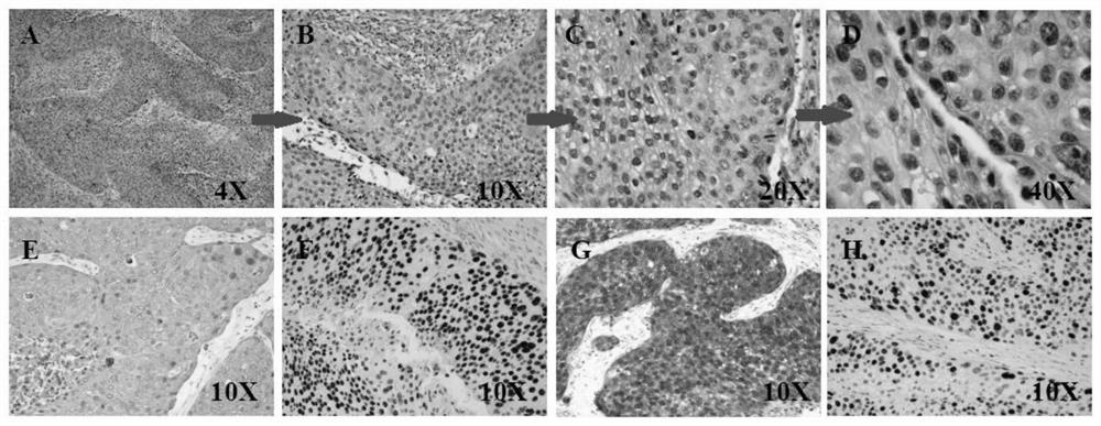
[0042] 1) Put the excised tissue block into the pre-cooled culture medium containing DMEM / F12 + 10% fetal bovine serum + 1% antibiotics, and store it at 4 °C. The HE staining test results of cervical squamous cell carcinoma tissue are as follows: image 3 As shown, A, B, C and D are HE staining of cervical squamous cell carcinoma, E is mouse anti-human AE1 / AE3 antibody staining (10X), I is mouse anti-human P63 antibody staining (10X), G is Mouse anti-human P16 antibody staining (10X), H is mouse anti-human Ki67 antibody staining (10X), it is clearly cervical squamous cell carcinoma, the proportion of cervical squamous cell carcinoma tissue components is as follows Figure 4As shown, cervical cancer tissue mouse anti-human AE1 / AE3, rabbit anti-human Vimentin and DAPI nuclear co-staining;

[0043] 2) Gently wipe the blood of the tissue block with gauze in the ultra-clean bench, rinse with PBS+10% antibiotics, put the cleaned tissue block into a 6cm petri dish with 0.5mL of DMEM-...


PUM


Abstract

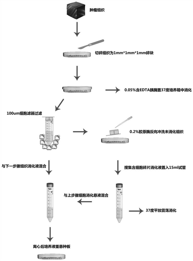
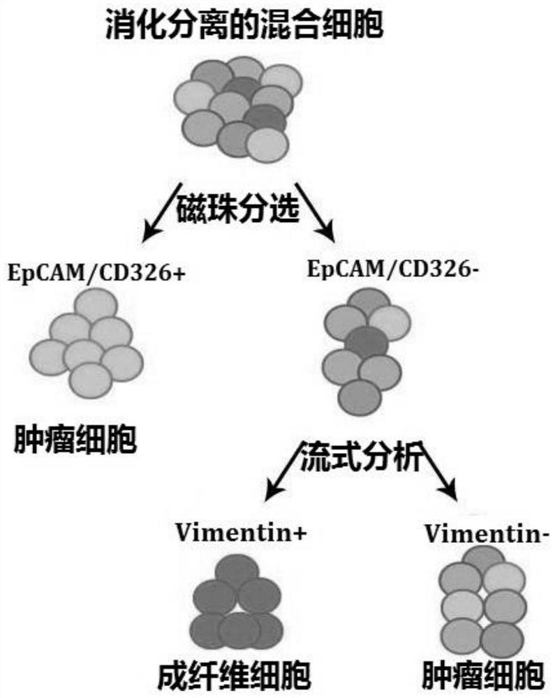
The invention discloses a method for separating and culturing primary cells of cervical cancer tumor, comprising the following steps: 1) placing tissue blocks in a pre-cooled sterile culture solution for processing; 2) cleaning and shredding the tissue; 3) digesting the tissue ; 4) Filter through a sterile cell filter to obtain the digestion filtrate and place it at a low temperature; 5) Rinse the sterile cell filter with collagenase and collect it into a centrifuge tube; 6) Place the centrifuge tube flat in a shaking box and shake well; 7) Combine 4) Step 6) Digestion solution, centrifuge, discard the supernatant; 8) Fully resuspend the cell pellet by pipetting; 9) Separation and passage; 10) Magnetic bead sorting. The beneficial effects of the invention are as follows: it can prolong the in vitro time of the tissue, improve the cell growth efficiency, shorten the time period, and have high purification efficiency, and provide a new and efficient scheme for the basic research of cervical cancer.

Description

technical field [0001] The invention relates to the field of modern biotechnology, in particular to a method for separating and culturing primary cells of cervical cancer tumors. Background technique [0002] Worldwide, cervical cancer (CC) is the second most common female malignant tumor after breast cancer. It is well known that persistent infection of high-risk HPV is the causative factor. Reduce the prevalence of CIN≥III by at least 50% (American Society of Clinical Oncology report, 2016), however, due to differences in mass screening and primary prevention coverage, and the lack of an effective HPV+ regression strategy, cervical cancer is still the most important of female reproductive system tumors. According to an article by the National Cancer Center of China (Cancer statistics in China, 2015), the incidence of cervical cancer is 99 / 100,000 people, and 31 / 100,000 people die, which is a serious threat to the physical and mental health of women in my country. Research...

Claims


Application Information

Patent Timeline
Patent Type & Authority Patents(China)
IPC IPC(8): C12N5/09
CPCC12N5/0693C12N2509/00
Inventor 吴令英张媛媛安菊生李宁李楠黄曼妮孙阳春王文鹏
Owner CANCER INST & HOSPITAL CHINESE ACADEMY OF MEDICAL SCI
Learn More