Abdominal ultrasound diagnostic equipment in general surgery department and use method thereof

An ultrasonic diagnosis and general surgery technology, applied in the directions of sonic diagnosis, ultrasonic/sonic/infrasonic diagnosis, infrasonic diagnosis, etc., can solve the problems of poor segmentation accuracy of ultrasonic images, high noise in low-frequency ultrasonic images, and reduced image accuracy, and achieve segmentation. High precision, fast speed, and good segmentation edge smoothness

Pending Publication Date: 2019-04-16
王立坤
View PDF0 Cites 1 Cited by
  • Summary
  • Abstract
  • Description
  • Claims
  • Application Information

AI Technical Summary

Problems solved by technology

[0004] (1) Low-frequency ultrasonic images collected by existing ultrasonic diagnostic equipment have large noise and low image quality; at the same time, the segmentation accuracy and smoothness of ultrasonic images are poor, which affects diagnosis
[0005] (2) In the process of collecting abdominal image data through the ultrasonic probe in the prior art, the acquired image is easily affected by the shadow effect and boundary noise on the detection area, which reduces the accuracy of image acquisition
[0006] (3) In the prior art, in the process of storing and collecting abdominal image data through memory, the image data needs to be compressed. During the compression process, the image will be distorted to a certain extent, reducing the clarity of the image.

Method used


Image

  • Abdominal ultrasound diagnostic equipment in general surgery department and use method thereof
  • Abdominal ultrasound diagnostic equipment in general surgery department and use method thereof
  • Abdominal ultrasound diagnostic equipment in general surgery department and use method thereof

Examples


Embodiment Construction

[0050]In order to further understand the content, features and effects of the present invention, the following examples are given, and detailed descriptions are given below with reference to the accompanying drawings.

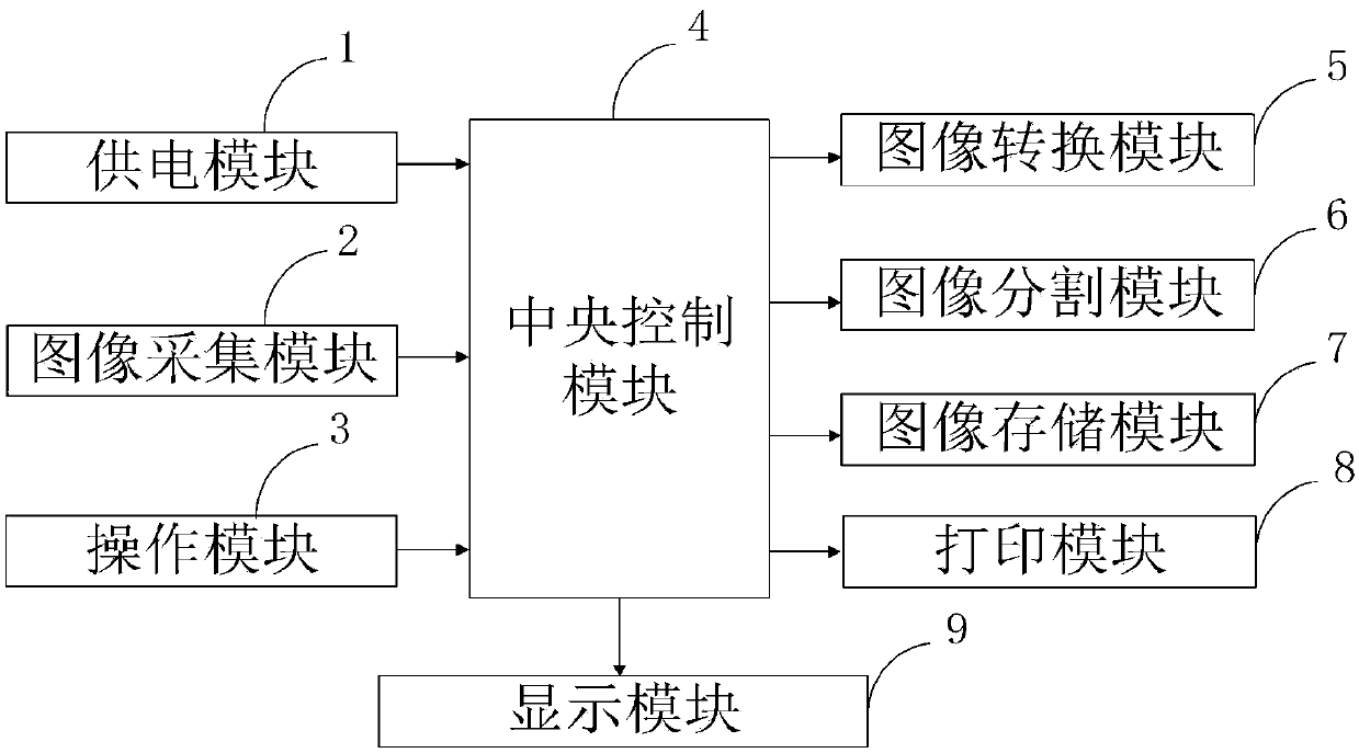
[0051] The structure of the present invention will be described in detail below in conjunction with the accompanying drawings.

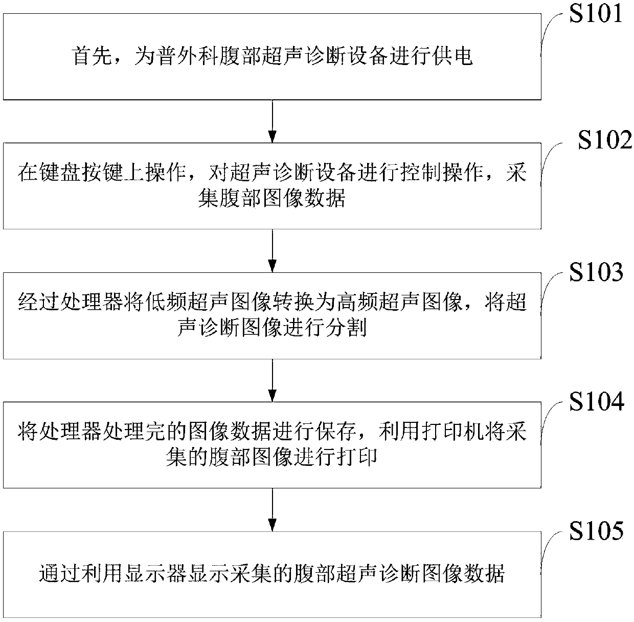
[0052] Such as figure 1 As shown, the method for using the abdominal ultrasonic diagnostic equipment for general surgery provided by the present invention comprises the following steps:

[0053] S101: First, supply power to the abdominal ultrasonic diagnostic equipment in general surgery;

[0054] S102: Operate on the keyboard keys to control and operate the ultrasonic diagnostic equipment, and collect abdominal image data;

[0055] S103: converting the low-frequency ultrasonic image into a high-frequency ultrasonic image through the processor, and segmenting the ultrasonic diagnostic image;

[0056] S104: Save the image data proce...


PUM


Abstract

The invention belongs to the technical field of ultrasound diagnosis, and discloses abdominal ultrasound diagnostic equipment in the general surgery department and a use method thereof. The abdominalultrasound diagnostic equipment in the general surgery department includes a power supply module, an image acquisition module, an operation module, a central control module, an image conversion module, an image segmentation module, an image storage module, a print module and a display module. The image conversion module is used for training an image control group of the same object at different frequencies to obtain the neural network parameter used for ultrasonic image conversion, so that an ultrasonic system generates an image high in speed, high in imaging quality, large in depth, high in resolution and low in noise. Meanwhile, the image segmentation module overcomes the deficiency of intelligent segmentation scenes applied to ultrasound image defects in a traditional image segmentationmethod, and the equipment is not sensitive to the complex and changeable high noise of the ultrasound image, has high segmentation accuracy, strong robustness and generalization, good smoothness of the segmentation edge, and is conducive to medical detection.

Description

technical field [0001] The invention belongs to the technical field of ultrasonic diagnosis, and in particular relates to an abdominal ultrasonic diagnosis device for general surgery and a method for using the same. Background technique [0002] General surgery refers to general surgery. In addition to general surgery, general general hospital surgery also includes orthopedics, neurosurgery, cardiothoracic surgery, and urology. Department of general surgery is a clinical discipline that treats liver, biliary tract, pancreas, gastrointestinal, anorectal, vascular diseases, thyroid and breast tumors, trauma and other diseases with surgery as the main method. It is the largest specialty in the surgical system. Ultrasonic diagnosis (ultrasonic diagnosis) is a diagnostic method that applies ultrasonic detection technology to the human body, understands the data and shape of physiological or tissue structures, discovers diseases, and makes prompts. Ultrasound used for medical dia...

Claims


Application Information

Patent Timeline
Patent Type & Authority Applications(China)
IPC IPC(8): A61B8/00G06T7/00G06T7/10
CPCA61B8/4411A61B8/5207G06T7/0012G06T7/10G06T2207/10132G06T2207/20081
Inventor 王立坤武雪亮屈明
Owner 王立坤
Why Eureka
Try Eureka