Extracellular vesicle and preparation method and parting analysis method thereof

An analysis method and cell technology, applied in cell dissociation methods, biochemical equipment and methods, animal cells, etc., can solve problems such as loss of information, detection of extracellular vesicles and specific cells, etc., to increase drug concentration and speed Effect

Pending Publication Date: 2019-04-19
WENZHOU MEDICAL UNIV
View PDF2 Cites 3 Cited by
  • Summary
  • Abstract
  • Description
  • Claims
  • Application Information

AI Technical Summary

Problems solved by technology

Most of the existing technologies use cracking methods to obtain extracellular vesicles, which cannot detect the degree of binding of extracellular vesicles to specific cells while ensuring the activity of extracellular vesicle surface molecules, and may lose a lot of information

Method used


Image

  • Extracellular vesicle and preparation method and parting analysis method thereof

Examples


Embodiment 1

[0026] Embodiment 1, the present invention provides a method for increasing the uptake rate of extracellular vesicles, comprising the following steps:

[0027] Step 1. Extraction of HCT116 extracellular vesicles by differential centrifugation: add the sample to a centrifuge tube, centrifuge at 500g for 15 minutes, and take the supernatant. Transfer the supernatant to a new centrifuge tube, centrifuge at 2000g for 15 minutes. Transfer the supernatant to a new centrifuge tube and centrifuge at 10,000g for 15 minutes; transfer the supernatant to an ultracentrifuge tube and centrifuge at 120,000g for 2 hours, remove the supernatant, collect the precipitate, and resuspend the pellet with PBS, which is the resuspension of extracellular vesicles liquid. Extracellular vesicle resuspension was quantified by BCA protein quantification kit;

[0028] Step 2: Add 1000 units of PNGFase according to 1 ug of extracellular vesicles, mix well, and incubate at 37°C for 2 hours;

[0029] Step ...

Embodiment 2

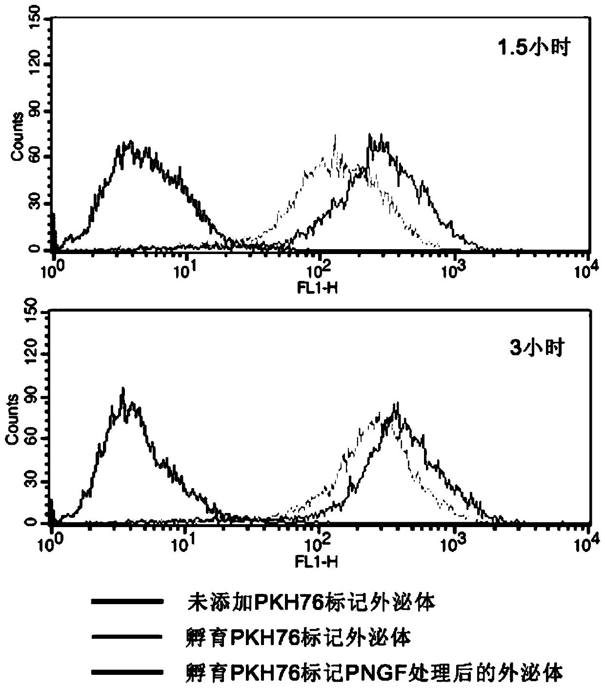
[0030] Example 2, the present invention provides an extracellular vesicle typing analysis method based on the uptake rate. The extracellular vesicles prepared according to Example 1 are used as the experimental group and HCT116 extracellular vesicles that have not been treated with PNGF As a control group, to determine the binding rate of two kinds of extracellular vesicles and HCT116 tumor cells, including the following steps:

[0031] Step 1. Take 20ug of extracellular vesicles from the experimental group and the control group. The extracellular vesicles of the two groups are labeled with PKH67 dyes respectively. After the reaction is terminated by adding 3% BSA, the excess PKH67 dye is removed by ultracentrifugation , resuspend extracellular vesicles with 100 μL PBS;

[0032] Step 2. Add an equal amount of different extracellular vesicles marked in step 1 to the HCT116 cell culture medium, and incubate with the HCT116 cells at 37°C and 5% carbon dioxide for 1.5h or 3h;

[...


PUM

PropertyMeasurementUnit
particle diameteraaaaaaaaaa

Abstract

The invention provides an extracellular vesicle and a preparation method and a parting analysis method thereof. Surface N-carbohydrate of the extracellular vesicle is removed, so that the taking-in rate of the extracellular vesicle relative to specific cells is increased. According to the method, by adopting co-culture of a living cell and the extracellular vesicle, through a fluorescent mark anda flow cytometry, cells, with fluorescence, in cell suspensions of different cell culture vessels and the fluorescence intensity are detected, and through the interaction relation of the living cell and the extracellular vesicle, parting analysis verification is conducted on the extracellular vesicle; in addition, the invention provides the extracellular vesicle which is prepared to be more efficiently taken in by HCT116.

Description

technical field [0001] The invention relates to the field of extracellular vesicles, in particular to an extracellular vesicle, a preparation method thereof, and a typing analysis method. Background technique [0002] Extracellular vesicles (EVs) are nanometer-sized, bilayer spherical proteoliposomes. Its particle size is about 50-100nm. The outer surface of the outer membrane vesicle is surrounded by a phospholipid bilayer, which is rich in water-soluble proteins, mRNA and microRNA and other substances. [0003] In terms of application to drug carriers, traditional drug carriers have large particle size and toxicity, relatively complex synthesis, and are easily phagocytized by macrophages, etc., and the carriers cannot effectively reach the tumor site. Traditional drug carriers include cationic liposomes and cationic polymers. Due to the presence of permanent positive charges on their surfaces, they can be combined with drugs through electrostatic interactions. However, a...

Claims


Application Information

Patent Timeline
Patent Type & Authority Applications(China)
IPC IPC(8): C12N5/09C12Q1/02G01N21/64A61K9/127A61K47/46
CPCA61K9/1271A61K47/46C12N5/0693C12N2509/00G01N21/6486G01N33/5005
Inventor 邓载安
Owner WENZHOU MEDICAL UNIV
Why Eureka
Try Eureka