Human-computer interaction type intracranial electrode positioning method and system based on three-dimensional convolution

A technology of three-dimensional convolution and positioning method, applied in the field of basic research of brain science, can solve the problems of time-consuming, three-dimensional image noise interference, and error-prone, and achieve the effect of simple and fast algorithm implementation, efficient and accurate positioning, and convenient manual marking.

Active Publication Date: 2019-04-23
NAT UNIV OF DEFENSE TECH
View PDF1 Cites 12 Cited by
  • Summary
  • Abstract
  • Description
  • Claims
  • Application Information

AI Technical Summary

Problems solved by technology

Since there are usually dozens or even hundreds of implanted electrodes, its position in three-dimensional space is often difficult to accurately correspond to the sketch set before operation. Numbering is not only time-consuming, but also due to problems such as overlapping of 3D images and noise interference, there may be problems such as mislabeling or wrong numbering, which will lead to directional errors in subsequent analysis, which has a great impact on clinical or scientific research
When marking electrode signals in 3D brain images, it is time-consuming, labor-intensive and error-prone to simply use manual methods. Since the positions of electrodes cannot be digitized when drawing the electrode sketches in the initial stage, the human-computer interaction method combining computer recognition and manual recognition can be used immediately. Improve the efficiency of marking while ensuring the reliability of the results, which is of great significance for real-time monitoring of abnormal discharge activities and accurate positioning of abnormal discharge computer areas

Method used


Image

  • Human-computer interaction type intracranial electrode positioning method and system based on three-dimensional convolution
  • Human-computer interaction type intracranial electrode positioning method and system based on three-dimensional convolution
  • Human-computer interaction type intracranial electrode positioning method and system based on three-dimensional convolution

Examples


Embodiment Construction

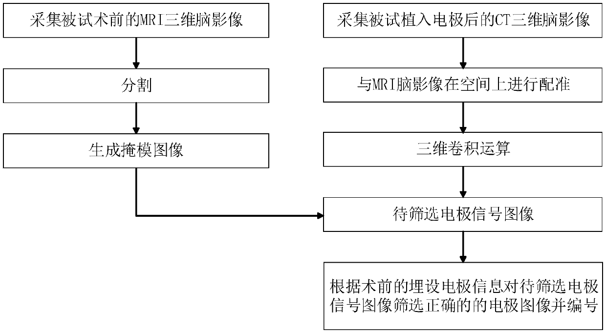
[0036] like figure 1 As shown, the implementation steps of the human-computer interactive intracranial electrode positioning method based on three-dimensional convolution in this embodiment include:

[0037] 1) Acquisition of preoperative MRI three-dimensional brain images and CT three-dimensional brain images after electrode implantation, and spatial registration of CT brain images and MRI brain images;

[0038]2) In the individual space, the MRI brain image is divided into five parts: gray matter, white matter, cerebrospinal fluid, dura mater and skull, and the three regions of gray matter, white matter and cerebrospinal fluid or the four regions of gray matter, white matter, cerebrospinal fluid and dura mater are combined as The target area of ​​the detection electrode is used as a mask image; the three-dimensional convolution operation is performed on the registered CT brain image to specifically distinguish the image signal of the intracranial electrode from other bright ...


PUM


Abstract

The invention discloses a human-computer interaction type intracranial electrode positioning method and system based on three-dimensional convolution. The method comprises the following steps: an MRIbrain image is segmented to generate a target area mask image of a detection electrode by combining an MRI three-dimensional brain image before an operation and a CT three-dimensional brain image after the operation; three-dimensional convolution operation is carried out on the registered CT brain image, the target area of the detection electrode is extracted according to the mask image and electrode signal images to be screened are extracted; and the electrode signal images to be screened are screened according to pre-operation embedded electrode information to obtain correct electrode imagesand numbering by man-machine interaction. According to the invention, the distinguishing capability of the electrode images is improved through the convolution operation, the distinguishing degree ofthe intracranial electrode in the CT brain image is effectively improved, the automatic identification of a computer is facilitated, and electrode screening and numbering are carried out through a high-efficiency man-machine interaction means, accurately positioning the coordinates of the intracranial electrode in the human brain is facilitated and the method has the advantages of simple principle, convenient achievement and stable result.

Description

technical field [0001] The present invention relates to the field of basic brain science research, in particular to a three-dimensional convolution-based human-computer interactive intracranial electrode positioning method and system, which are used to locate the subject's brain using preoperative MRI three-dimensional brain images and postoperative CT three-dimensional brain images. Precise placement of intracranial electrodes. Background technique [0002] In recent years, brain science and cognitive science have developed rapidly, and research on various aspects of human brain and cognition is constantly making breakthroughs. In order to better monitor the neural activity of the brain, Positron Emission Computed Tomography-Computed Tomography (PET-CT) technology, functional magnetic resonance technology (fMRI), electroencephalogram (EEG) ), synchronous EEG-functional magnetic resonance (EEG-fMRI) and other imaging or signal detection techniques have been used in in-depth...

Claims


Application Information

Patent Timeline
Patent Type & Authority Applications(China)
IPC IPC(8): A61B34/20A61B5/04
CPCA61B34/20A61B2034/2065A61B5/24
Inventor 曾令李苏建坡胡德文沈辉汤鹏飞
Owner NAT UNIV OF DEFENSE TECH
Why Eureka
Try Eureka