A pulmonary nodule automatic detection method and system based on a pulmonary CT sequence

A technology for automatic detection of pulmonary nodules, applied in image data processing, instruments, calculations, etc., to achieve the effect of reducing difficulty, high detection rate, and enhancing fitting ability

Active Publication Date: 2019-04-26
心医国际数字医疗系统(大连)有限公司
View PDF7 Cites 17 Cited by
  • Summary
  • Abstract
  • Description
  • Claims
  • Application Information

AI Technical Summary

Problems solved by technology

[0013] In order to solve the problem of improving the network fitting ability and the detection rate of candidate nodules, the present invention proposes an automatic detection method for pulmonary nodules based on lung CT sequences, using a 3D model with a residual structure and a feature map structure The full convolutional network is used to obtain candidate nodules, and a 3D convolutional network with multi-model fusion is used to remove false positives

Method used


Image

  • A pulmonary nodule automatic detection method and system based on a pulmonary CT sequence
  • A pulmonary nodule automatic detection method and system based on a pulmonary CT sequence
  • A pulmonary nodule automatic detection method and system based on a pulmonary CT sequence

Examples


Embodiment Construction

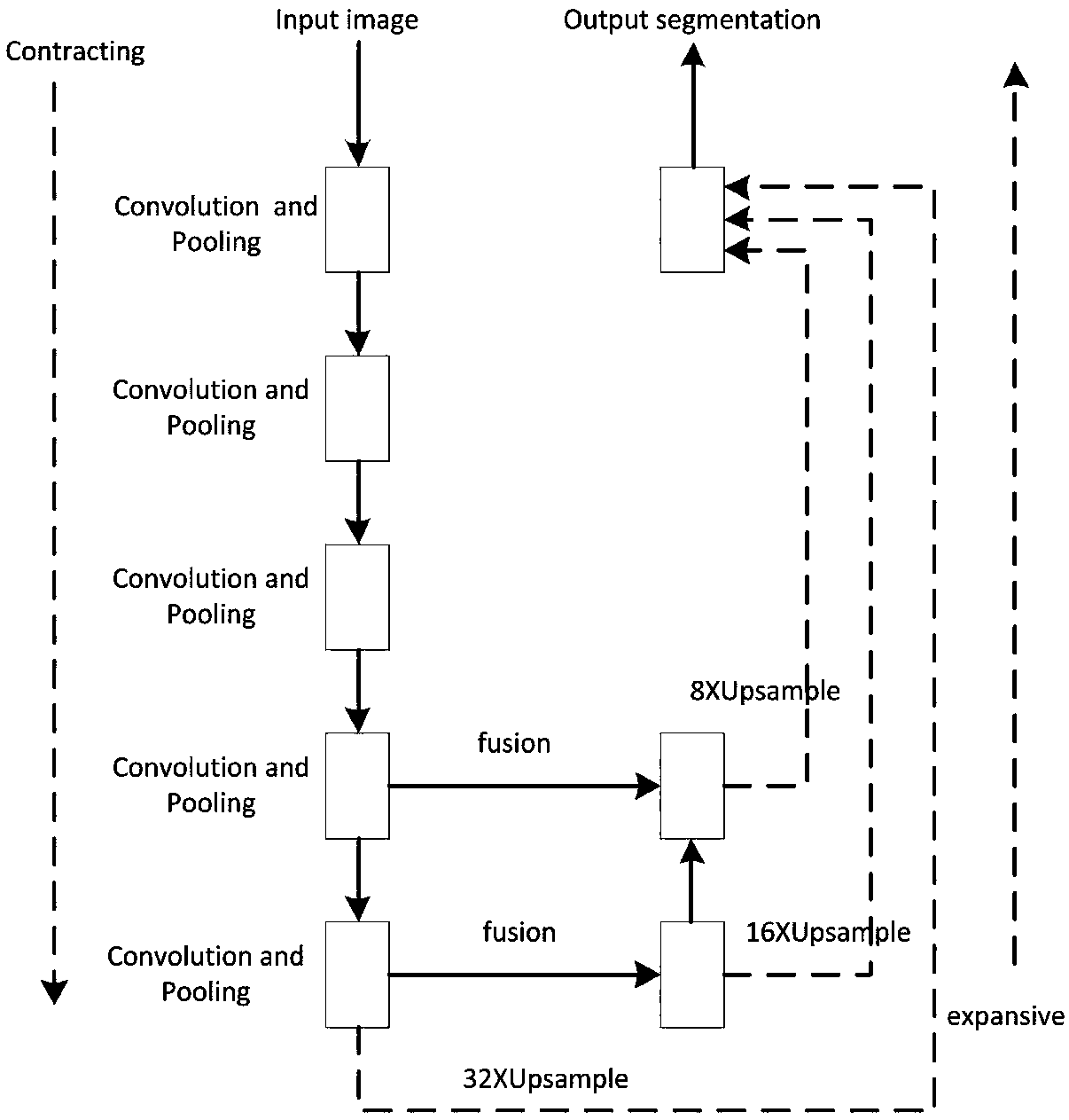
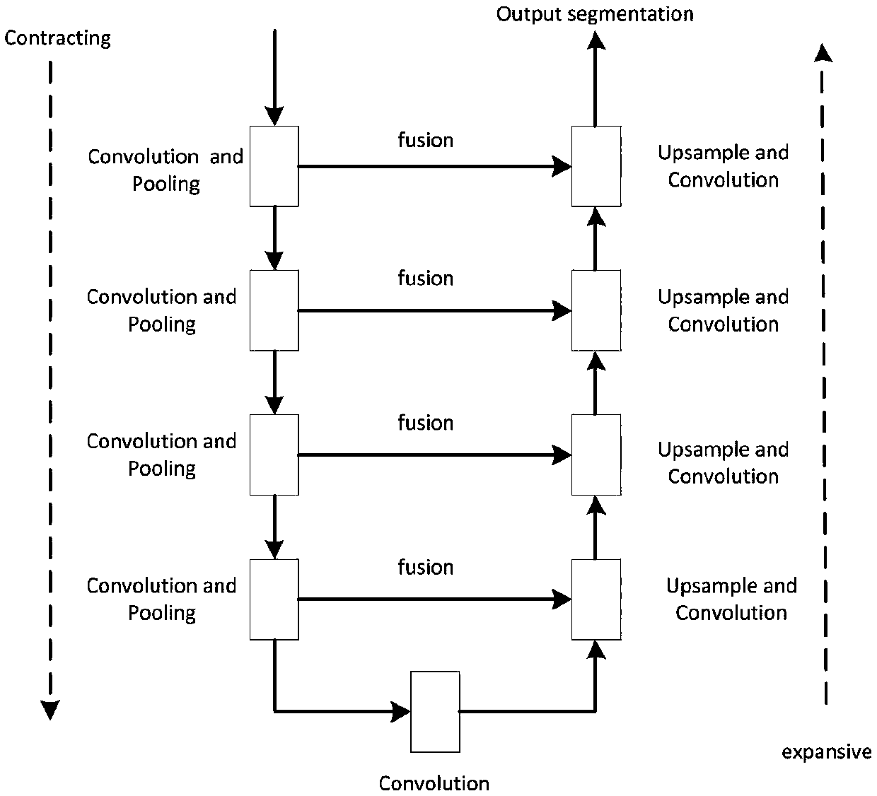
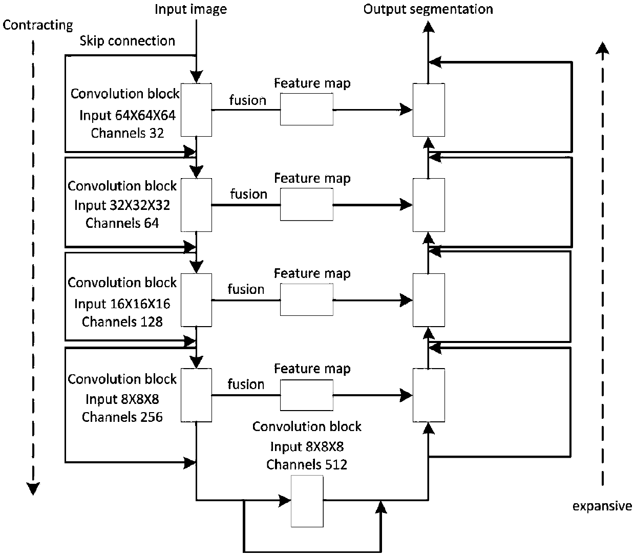
[0041] An automatic detection method for pulmonary nodules based on lung CT sequences. This detection method has two stages: candidate nodule acquisition and false positive removal. It needs to train a full convolutional network and a multi-model fusion 3D convolutional network respectively, and use the trained model is tested.

[0042] That is, the detection method includes:

[0043] S1. Data preprocessing;

[0044] S2. Screen candidate nodules using a fully convolutional network;

[0045] S3. Using an image processing method to change the probability map into the coordinates of the center point of the nodule and the radius;

[0046] S4. Using multi-model fusion 3D convolutional network detection to obtain the final determined nodule coordinates and corresponding radius sets.

[0047] Wherein: step S2 involves using a full convolutional network to screen candidate nodules, and the full convolutional network used in this step needs to be trained to achieve detection, and th...


PUM


Abstract

The invention discloses a pulmonary nodule automatic detection method and system based on a pulmonary CT sequence, belongs to the field of medical image segmentation, and aims to improve the network fitting capability and the candidate nodule detection rate. The method comprises steps of S1, data preprocessing; S2, screening candidate nodules by using a full convolutional network; S3, using an image processing method to change the probability graph into nodule center point coordinates and radiuses; And S4, using multi-model fused 3D convolutional network detection to obtain a finally determined nodule coordinate and a corresponding radius set, better utilizing the spatial information of the lung CT, and introducing a residual structure and a feature mapping structure to enhance the fittingcapability of the 3D full convolutional network.

Description

technical field [0001] The invention belongs to the field of medical image segmentation, and relates to a lung nodule automatic detection method and system based on a lung CT sequence. Background technique [0002] Lung cancer is a disease with high mortality rate and high incidence frequency. Survival for lung cancer is highly correlated with the stage of the disease when it is first diagnosed. Because most of the early lung cancer has no obvious symptoms, the clinical diagnosis of lung cancer is often in the middle and late stage. Not only the treatment cost is high, but also the treatment effect is not ideal. Therefore, early detection and early diagnosis of lung cancer is very necessary. Lung cancer often manifests as nodules in the early stage, so the detection and diagnosis of pulmonary nodules are of great significance to the early detection of lung cancer. CT has high tissue resolution and is an ideal tool for the detection and diagnosis of pulmonary nodules. Wit...

Claims


Application Information

Patent Timeline
Patent Type & Authority Applications(China)
IPC IPC(8): G06T7/00G06T7/11G06T7/62
CPCG06T7/0012G06T7/11G06T7/62G06T2207/10081G06T2207/20076G06T2207/20081G06T2207/20084G06T2207/30064
Inventor 王兴维邰从越刘龙尹延伟王慧史黎鑫刘慧芳
Owner 心医国际数字医疗系统(大连)有限公司
Why Eureka
Try Eureka