Humanized adipose-derived stem cell exosome and preparation method and application thereof

A technology of fat stem cells and exosomes, applied in the field of stem cells and biomedicine

Pending Publication Date: 2019-05-14
博品(上海)生物医药科技有限公司
View PDF5 Cites 7 Cited by
  • Summary
  • Abstract
  • Description
  • Claims
  • Application Information

AI Technical Summary

Problems solved by technology

However, it is unknown what kind of tissue repair and regeneration exosomes from different stem cell sources or different exosomes from the same stem cell source are suita

Method used


Image

  • Humanized adipose-derived stem cell exosome and preparation method and application thereof
  • Humanized adipose-derived stem cell exosome and preparation method and application thereof
  • Humanized adipose-derived stem cell exosome and preparation method and application thereof

Examples


Embodiment 1

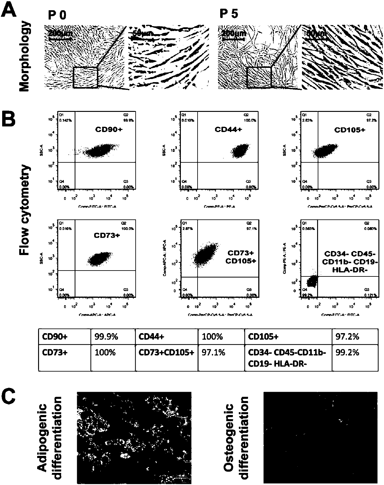
[0125] Example 1. Isolation and identification of hASC

[0126] The hASCs isolated by the present inventors maintain fibroblast-like morphology from the P0 generation, are uniform in size and closely arranged, and grow in a spiral or radial shape. There was no significant change in the shape, size and density of the P5 generation cells ( figure 1 A). The results of flow cytometry showed that protein markers such as CD73 (100%), CD44 (100%), CD90 (99.9%), and CD105 (97.2%) were highly expressed on the surface of hASCs, and hematopoietic cell line molecular markers CD34 and CD45 were hardly expressed , CD19, etc. (0.8%), confirming that the isolated hASCs are mesenchymal stem cells with high purity ( figure 1 B). After cultured in adipogenic medium for 3 weeks, a large number of transparent lipid droplets with good refraction properties can be seen in the cytoplasm under the microscope, which turns red after staining with Oil Red O; after cultured in osteogenic medium for 3 w...

Embodiment 2

[0127] Example 2. Extraction and identification of hASC exosomes

[0128] The hASC exosomes were extracted and identified according to "Materials and Methods", and the results are as follows:

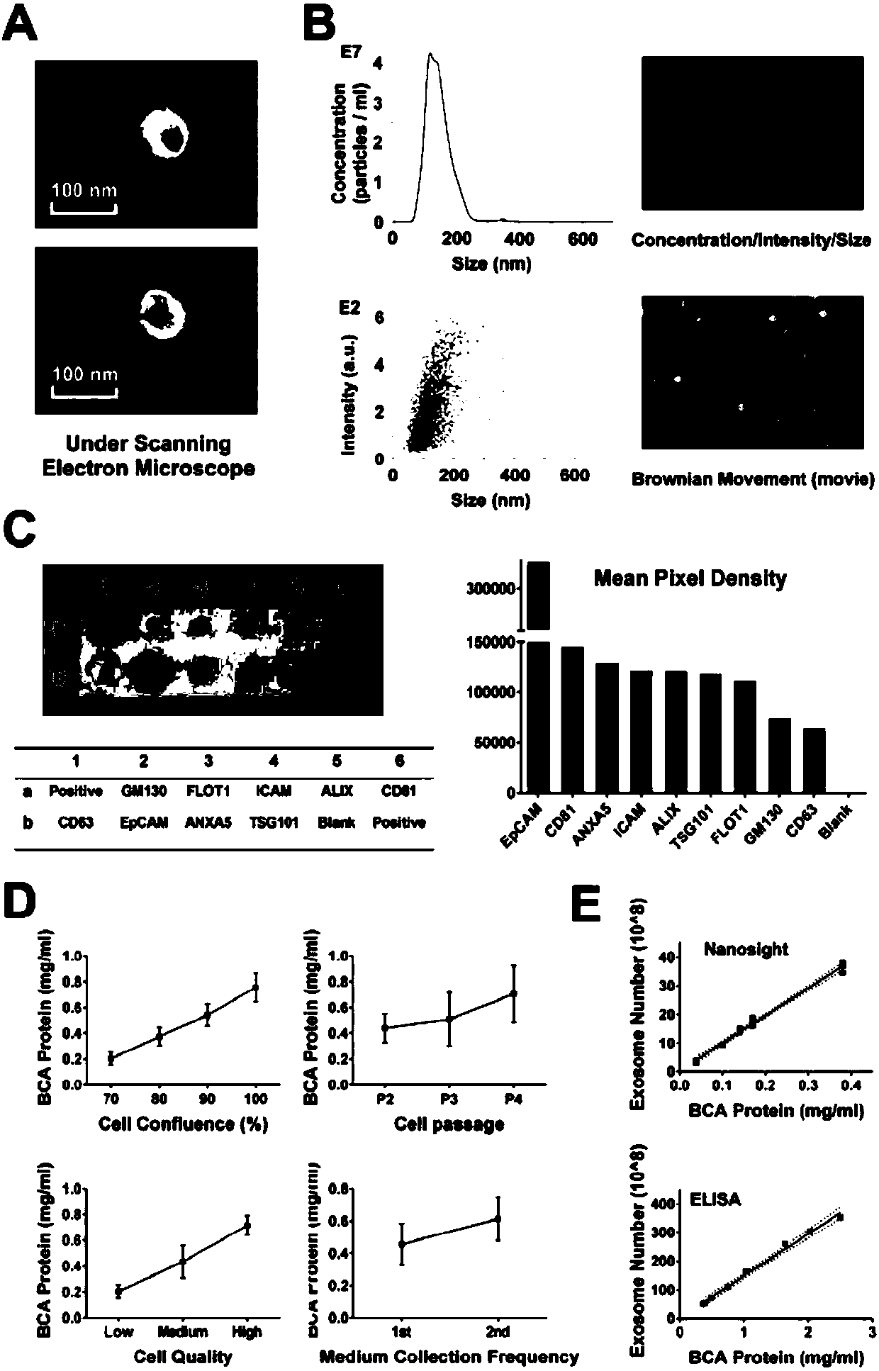
[0129] 1. Scanning electron microscope detection: Under the scanning electron microscope, it can be seen that hASC exosomes have a saucer-like or concave hemispherical structure of uniform size, with obvious membrane structure, clear three-dimensional structure, and the size is about 30-200nm ( figure 2 A).

[0130] 2. Nanosight particle size analyzer detection: Nanosight particle size analyzer detects hASC exosomes. From the size / concentration curve, it can be seen that the size of hASC exosomes is about 30-200nm, and the average size is about 100nm. The measured sample concentration is about 4×10 7 Particles / ml. From the size / intensity curve, it can be seen that the intensity of hASC exosomes is about 0.5-6a.u., and the intensity of particles with a size of about 150nm is the highe...

Embodiment 3

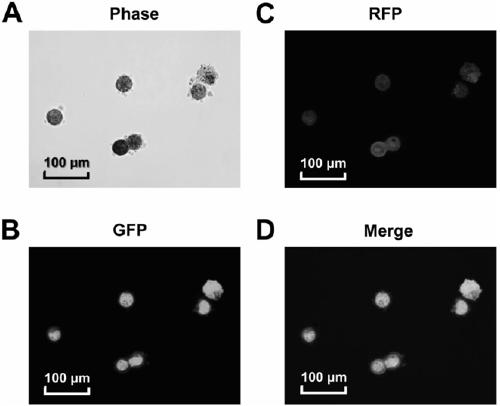
[0133] Example 3. In vitro uptake of hASC exosomes by hepatocytes

[0134] According to "Materials and Methods", the in vitro uptake of hASC exosomes by hepatocytes was tested, and the results are as follows:

[0135] After hASC exosomes were incubated with hepatocytes for 2 h ( image 3 A), it can be seen that red and green fluorescent particles appear in the cytoplasm of liver cells at the same time, and the green fluorescent particles are proteins in exosomes ( image 3 B), red fluorescence labeled RNA in exosomes ( image 3 C), and no decrease in fluorescence was observed over time. It shows that hASC exosomes can penetrate the liver cell membrane, enter the liver cytoplasm, and exist in the liver cells for a long time.


PUM

PropertyMeasurementUnit
Sizeaaaaaaaaaa
Sizeaaaaaaaaaa

Abstract

The invention provides an exosome from adipose-derived stem cells and a preparation method thereof. The exosome from the adipose-derived stem cells can be used for treating an acute hepatic failure, liver fibrosis, liver cirrhosis and other liver diseases. The exosome from the adipose-derived stem cells can remarkably increase the survival rate of a patient with the acute hepatic failure and evencan realize the 100% survival rate of the patient with the acute hepatic failure under high concentration.

Description

technical field [0001] The invention relates to the fields of stem cells and biomedicine. Specifically, the present invention relates to human-derived adipose stem cell (Human-Adipose Derived Stem Cell, hADSC) exosomes and its preparation method and application in the treatment of acute liver failure and other diseases. Background technique [0002] Liver failure is a serious threat to human health. Orthotopic liver transplantation is currently the most effective treatment, but it is difficult to be used as a common treatment for patients with end-stage liver disease because of its high cost and limitation of liver source[1-3]. With the maturity of stem cell (Stem cell) technology, it has been confirmed that stem cells can promote the regeneration of damaged organ tissue cells through "cell replacement" or "paracrine effect"[4-7]. Adipose stem cells (Human adipose-derived stem cells, hASC), as a kind of adult stem cells, have higher proliferation rate and stronger differen...

Claims


Application Information

Patent Timeline
IPC IPC(8): C12N5/0775A61P1/16
Inventor 徐俊傅青春金银鹏高正良李洪超王晢
Owner 博品(上海)生物医药科技有限公司
Why Eureka
Try Eureka