An ultrasonic image blood vessel diameter automatic measurement method

An ultrasonic image and automatic measurement technology, applied in the field of medical image analysis, can solve problems such as unconsidered and low calculation accuracy

Active Publication Date: 2019-06-14
SHENZHEN COMEN MEDICAL INSTR
View PDF10 Cites 8 Cited by
  • Summary
  • Abstract
  • Description
  • Claims
  • Application Information

AI Technical Summary

Problems solved by technology

This method does not consider the influence of noise on the edge of the blood vessel, and

Method used


Image

  • An ultrasonic image blood vessel diameter automatic measurement method
  • An ultrasonic image blood vessel diameter automatic measurement method
  • An ultrasonic image blood vessel diameter automatic measurement method

Examples


Embodiment Construction

[0028] In order to make the objectives, technical solutions and advantages of the present invention clearer, the following further describes the present invention in detail with reference to the accompanying drawings and embodiments. It should be understood that the specific embodiments described herein are only used to explain the present invention, but not to limit the present invention.

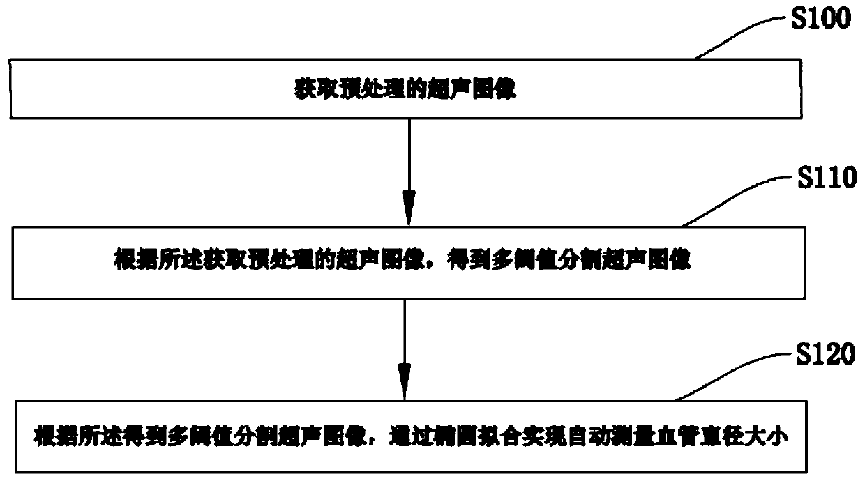
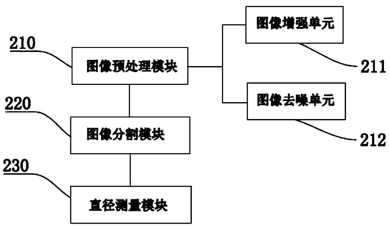
[0029] The embodiment of the invention discloses a method for automatically measuring the diameter of a blood vessel in an ultrasound image, such as figure 1 with image 3 Shown, including:

[0030] Step S110, acquiring a preprocessed ultrasound image;

[0031] Step S120: Obtain a multi-threshold segmented ultrasound image according to the acquired preprocessed ultrasound image;

[0032] In step S130, the ultrasound image is segmented according to the obtained multi-threshold value, and the blood vessel diameter is automatically measured through ellipse fitting.

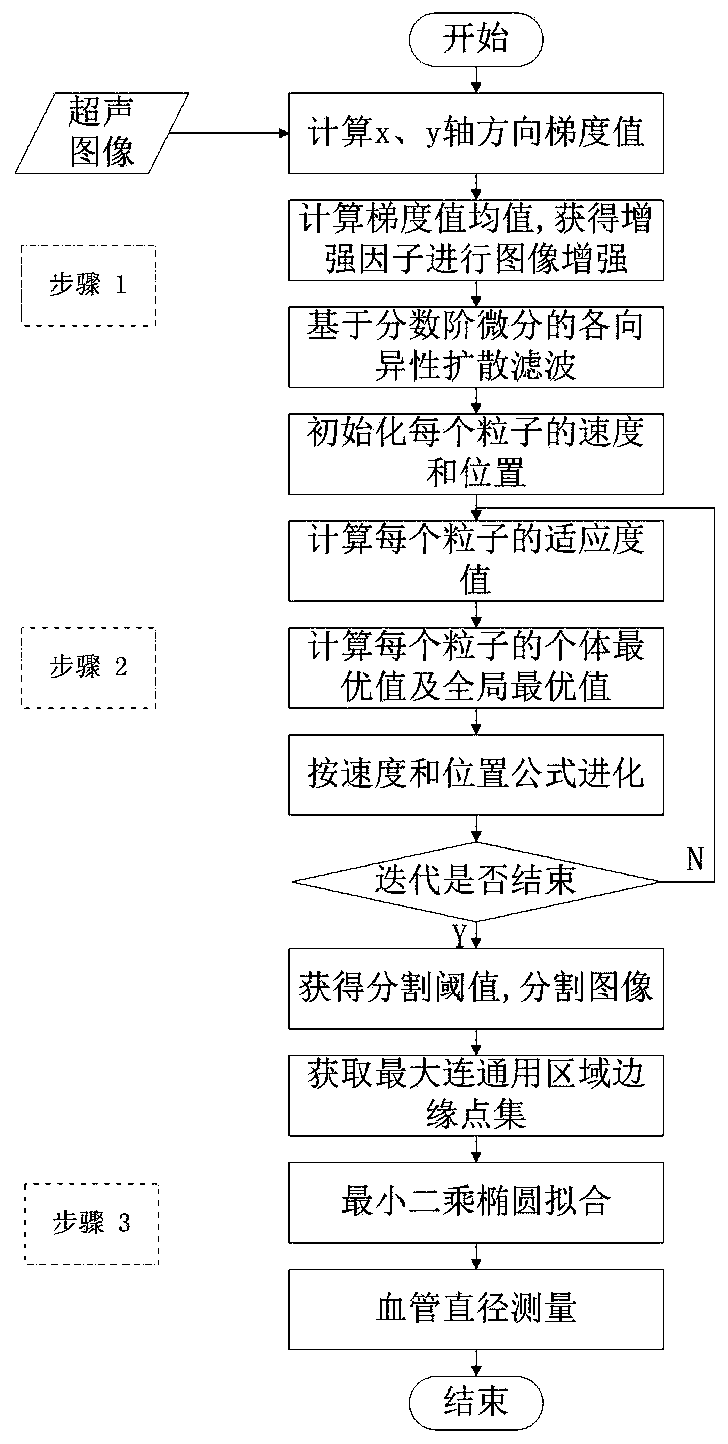
[0033] Such as Figure 4 As shown,...


PUM


Abstract

The invention relates to the technical field of medical image analysis, and discloses an ultrasonic image blood vessel diameter automatic measurement method, which comprises the following steps of obtaining a preprocessed ultrasonic image; obtaining a multi-threshold segmentation ultrasonic image according to the obtained pre-processed ultrasonic image; and automatically measuring the diameter ofthe blood vessel through ellipse fitting according to the obtained multi-threshold segmentation ultrasonic image. An ultrasonic image is enhanced, a large number of noise particles are distributed throughout the ultrasonic image, and by carrying out noise smoothing on the enhanced image, performing multi-threshold image segmentation on the smoothed image, and dividing the ultrasonic image into four different areas according to pixel gray values, due to the fact that the shape of the blood vessel is generally round, and the diseased blood vessel is generally oval, and by carrying out oval curvefitting on the segmented blood vessel area, the manual intervention is not needed in the whole algorithm, the automatic measurement of the diameter of the blood vessel in the ultrasonic image is achieved, and therefore an important clinical auxiliary diagnosis technology is provided for the PICC or CVC operation.

Description

Technical field [0001] The invention relates to the technical field of medical image analysis, in particular to a method for automatically measuring blood vessel diameters in ultrasound images. Background technique [0002] PICC (central venous catheterization via peripheral venous puncture) or CVC (central venous catheter), as a common clinical infusion tool, is a kind of infusion that can be punctured through the skin and stayed in the venous cavities (superior vena cava, inferior vena cava, brachiocephalic vein) , Internal jugular vein, subclavian vein, iliac vein, femoral vein) special silicone tube for long-term infusion. During the operation, if the diameter of the catheter is too large or too small, it will bring adverse consequences to the operation. [0003] In order to be able to select a suitable catheter for intubation according to the diameter of the blood vessel, it is necessary to use an ultrasound device to perform an imaging display of the blood vessel area at the...

Claims


Application Information

Patent Timeline
IPC IPC(8): G06T7/00G06T7/11G06T7/136G06T7/62
Inventor 李聪王兴红
Owner SHENZHEN COMEN MEDICAL INSTR
Why Eureka
Try Eureka