Method for positioning and guiding bone drilling through 3D modelling of medical image

A 3D modeling and medical imaging technology, applied in the medical field, can solve problems such as drilling too deep, dangerous, and tissue damage, and achieve the effect of improving accuracy and avoiding omissions

Inactive Publication Date: 2019-06-18
成都真实维度科技有限公司 +1
View PDF0 Cites 8 Cited by
  • Summary
  • Abstract
  • Description
  • Claims
  • Application Information

AI Technical Summary

Problems solved by technology

[0003] In orthopedic treatment, it is often necessary to drill the bone. However, the existing bone drilling method has some defects in the process of use. For example, it needs medical personnel to locate the drilling position through experience, and there is a possibility that the drilling may deviate.
For beginners or inexperienced medical personnel, the problem of drilling too deep often occurs during use, which will damage the tissue and is very dangerous

Method used


Image

  • Method for positioning and guiding bone drilling through 3D modelling of medical image
  • Method for positioning and guiding bone drilling through 3D modelling of medical image
  • Method for positioning and guiding bone drilling through 3D modelling of medical image

Examples


Embodiment 1

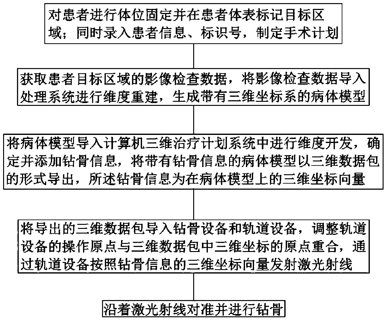
[0061] Such as figure 1 As shown, a method for positioning and guiding orthopedic drilling through three-dimensional modeling of medical images comprises the following steps:

[0062] Step A: Fix the patient's position and mark the target area on the patient's body surface; at the same time, enter the patient's information and identification number, and formulate the operation plan;

[0063] Step B: Obtain the image inspection data of the patient's target area, import the image inspection data into the processing system for dimensional reconstruction, and generate a patient model with a three-dimensional coordinate system;

[0064] Step C: Import the patient model into the computer 3D treatment planning system for dimension development, determine and add drill bone information, export the patient model with drill bone information in the form of a 3D data package, and the drill bone information is in the patient model The three-dimensional coordinate vector on ;

[0065] Step...

Embodiment 2

[0069] Such as figure 1 As shown, a method for positioning and guiding orthopedic drilling through three-dimensional modeling of medical images comprises the following steps:

[0070] Step A: Fix the patient's position and mark the target area on the patient's body surface; at the same time, enter the patient's information and identification number, and formulate the operation plan;

[0071] Step B: Obtain the image inspection data of the patient's target area, import the image inspection data into the processing system for dimensional reconstruction, and generate a patient model with a three-dimensional coordinate system;

[0072] Step C: Import the patient model into the computer 3D treatment planning system for dimension development, determine and add drill bone information, export the patient model with drill bone information in the form of a 3D data package, and the drill bone information is in the patient model The three-dimensional coordinate vector on ;

[0073] Step...

Embodiment 3

[0092] A method for positioning and guiding orthopedic drilling through three-dimensional modeling of medical images, comprising the following steps:

[0093] Step A: Fix the patient's position and mark the target area on the patient's body surface; at the same time, enter the patient's information and identification number, and formulate the operation plan;

[0094] Step B: Obtain the image inspection data of the patient's target area, import the image inspection data into the processing system for dimensional reconstruction, and generate a patient model with a three-dimensional coordinate system;

[0095] Step C: Import the patient model into the computer 3D treatment planning system for dimension development, determine and add drill bone information, export the patient model with drill bone information in the form of a 3D data package, and the drill bone information is in the patient model The three-dimensional coordinate vector on ;

[0096] Step D: Import the exported 3D...


PUM


Abstract

The invention belongs to the technical field of medical technologies and discloses a method for positioning and guiding bone drilling through 3D modelling of a medical image. The method comprises steps as follows: step A, the body position of a patient is fixed, and a target area is marked on the body surface of the patient; besides, information and identification number of the patient are recorded, and an operation plan is formulated; step B, image examination data of the target area of the patient are acquired and imported into a processing system for dimensional reconstruction; step C, a sick body model is imported into a computer 3D treatment plan system for dimension development, and information of to-be-drilled bone is determined and added; step D, an exported 3D data packet is imported into bone drilling equipment and track equipment, and an operation origin of the track equipment and an origin of a 3D coordinate in the 3D data packet coincide; step E, alignment along a laser ray and bone drilling are performed. When bone is required to be fixed, the bone drilling equipment is used for drilling the bone, later implantation of bone nails is facilitated, and the bone nail implantation accuracy is improved.

Description

technical field [0001] The invention belongs to the technical field of medical technology, and in particular relates to a method for positioning and guiding orthopedic drilling through three-dimensional modeling of medical images. Background technique [0002] With the development and progress of science and technology, orthopedics has made great progress in diagnosis and treatment. The establishment and development of hand surgery has rapidly popularized and improved the diagnosis and treatment of hand trauma; the extensive development of microsurgery has enabled the free reconstruction of polydactyly, the anastomosis and release of nerve bundles, and the anastomosis between small arteries of 0.2mm It is no longer a myth; using new surgical methods to treat scoliosis, cervical spondylosis and cervical stenosis and other diseases has achieved good orthopedic results; various effective surgical methods for arthritis patients have made bedridden patients Patients stand up aga...

Claims


Application Information

Patent Timeline
IPC IPC(8): A61B17/17A61B34/10A61B34/20A61B90/13
Inventor 赵德伟曲飞寰赵振华
Owner 成都真实维度科技有限公司
Why Eureka
Try Eureka