Spinal surgery minimally invasive surgery navigation system

A minimally invasive surgery and navigation system technology, applied in the field of spinal surgery minimally invasive surgery navigation system, can solve the problems of long time for intraoperative image registration, long time for accurate three-dimensional reconstruction, and inability to accurately position, so as to improve clinical practicability and Generalization, precise and stable control of puncture angle and depth, and the effect of avoiding damage to normal tissues

Active Publication Date: 2019-06-25
吕海 +2
View PDF10 Cites 14 Cited by
  • Summary
  • Abstract
  • Description
  • Claims
  • Application Information

AI Technical Summary

Problems solved by technology

The current navigation system for spinal surgery generally has the following problems that affect its clinical application and promotion: 1. The three-dimensional reconstruction of preoperative CT images fails to fully fuse MRI images that clearly show soft tissues such as intervertebral discs and nerves with CT images. Although it is possible to reconstruct the 3D images of bones, it is still not possible to accurately locate the positions of nerves, blood vessels, and intervertebral discs that compress nerves; 2. Preoperative 3D reconstruction takes a long time; 3. Preoperative 3D reconstruction and intraoperative image matching Long accurate time, poor clinical practicability
Therefore, the minimally invasive surgical navigation system for intervertebral disc herniation currently on the market needs to be further improved.

Method used


Image

  • Spinal surgery minimally invasive surgery navigation system
  • Spinal surgery minimally invasive surgery navigation system
  • Spinal surgery minimally invasive surgery navigation system

Examples


Embodiment 1

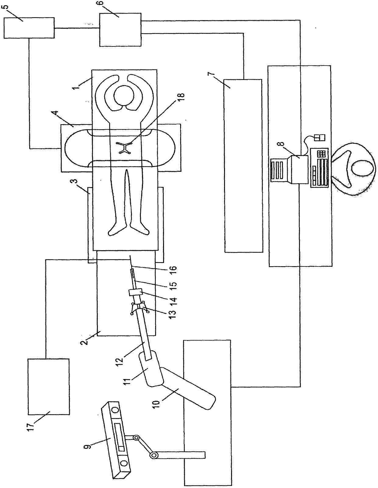
[0030] Embodiment one: if figure 1 , figure 2 , Figure 5 As shown in the figure, a navigation system for minimally invasive spinal surgery uses CT, MRI and other imaging equipment to obtain image data of the patient's spinal lesion area before the operation and inputs it into the system controller 8 for storage. The image processor 6 reads the image data of the system controller 8. The CT and MRI image information of the patient's preoperative spinal lesion is fused, the useful data is extracted for image segmentation, and the three-dimensional image model is reconstructed to display the patient's vertebrae, joints, nerves, blood vessels, and intervertebral disc tissue that compresses the nerve as a whole. Ligaments, preoperative surgical path planning based on the position of herniated disc and compressed nerve.

[0031] During the operation, the patient lies prone on the operating table 1, and the operating table 1 is connected to the lower base 3 through the slide rail as...

Embodiment 2

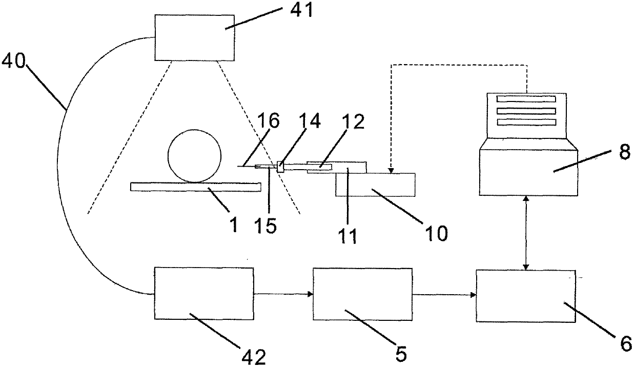
[0036] Embodiment two: if figure 1 , figure 2 , Figure 5 Shown, a kind of spinal surgery minimally invasive operation navigation system, this embodiment is compared with the first embodiment, the difference is: in this embodiment, the scanner 4 that is used to scan patient's spinal lesion is two-dimensional C Arm 40, the scanner 4 irradiates the patient with X-rays through the X-ray light source 41, and receives the X-ray information passing through the patient through the X-ray imaging device 42. After the X-rays penetrate the human body, the attenuation degree of different parts Different image information is obtained, and the image signal converter 5 converts the analog signal information obtained with different attenuation degrees into digital signal information, and the digital signal information is processed by the image processor 6 to obtain a two-dimensional bone image of the patient's spinal lesion, which is controlled by the system Under the control of the device...

Embodiment 3

[0037] Embodiment three: as figure 1 , Figure 5 , Figure 6 Shown, a kind of spinal surgery minimally invasive surgery navigation system, this embodiment is compared with the first embodiment, the difference is: in this embodiment, the scanner 4 that is used to scan patient's spinal lesion is O-shaped arm 43. After the X-ray penetrates the human body, the image information is obtained from the different attenuation degrees of different parts, and the image signal converter 5 converts the analog signal information obtained by the different attenuation degrees into digital signal information, and the digital signal information passes through the image processor 6 Computing and processing to obtain the two-dimensional bone image of the patient's spinal lesion. Under the control of the system controller 8, the image processor 6 performs three-dimensional reconstruction on the two-dimensional bone image, and the obtained three-dimensional reconstructed image is fused with the pat...


PUM


Abstract

The invention relates to a spinal surgery minimally invasive surgery navigation system. The spinal surgery minimally invasive surgery navigation system comprises an imaging portion, a treatment portion, and a navigation portion, wherein an imaging portion scanner performs scaning and imaging around a treatment object; the treatment portion includes a mechanical arm, a connecting rod, a power supply unit, a clipper, a puncture channel into a lesion of a spinal column, and an ablation electrode needle. The puncture channel is a minimally invasive surgical working cannula having a diameter of about 2 mm. The system has the advantages of simple structure, low cost and easy operation, and the CT and MRI images are combined to integrally reconstruct the intervertebral disc tissues of the patient's bone structure, nerves, blood vessels and compression nerves as well as the ligament before the surgery, according to the position of protrusion of intervertebral disc and the nerve compression, areasonable surgical approach is designed before the surgery, and the intraoperative scanning bone image is registered, a robot arm is remotely controlled to accurate ablate the compression nerve tissue through real-time navigation, and the safety and stability are good. The system avoids damage to normal tissues caused by doctor fatigue during surgery and effectively reduces the damage of radiation to patients and doctors.

Description

technical field [0001] The invention belongs to the field of medical instruments, in particular to a navigation system for minimally invasive spinal surgery. Background technique [0002] Minimally invasive surgery is the main development trend of surgical treatment of intervertebral disc herniation. At present, the surgical methods for intervertebral disc herniation require opening a window on the outer wall of the spinal canal of the diseased segment, and then through this window, the intervertebral disc, ligament, and hyperplastic calcified tissue that compress the nerve are removed. Depending on the location and size of the window on the outer wall of the spinal canal, normal tissues such as vertebrae or paravertebral bones, joints, ligaments, and muscles will be damaged to varying degrees. After surgery, it is easy to cause vertebral instability, nerve damage, and scar recurrence. Compressed nerves and other complications. [0003] During the process of resection of d...

Claims


Application Information

Patent Timeline
Patent Type & Authority Applications(China)
IPC IPC(8): A61B34/20A61B34/10A61B34/30
Inventor 吕海冯前进张伟聪王敏赵庆豪苏志海
Owner 吕海
Why Eureka
Try Eureka