Application of TBK1 as an E3 ubiquitin ligase

A ubiquitin ligase and virus technology, applied in the field of biomedicine, can solve the problems of strong virus virulence and incomplete virus inactivation.

Active Publication Date: 2020-05-15
LANZHOU INST OF VETERINARY SCI CHINESE ACAD OF AGRI SCI
View PDF1 Cites 0 Cited by
  • Summary
  • Abstract
  • Description
  • Claims
  • Application Information

AI Technical Summary

Problems solved by technology

However, due to unsafe factors such as strong virus virulence, incomplete virus inactivation, and live virus escape from processing plants, the outbreak of FMD in some parts of the world seems to be related to the residual live virus in the inactivated vaccine.

Method used


Image

  • Application of TBK1 as an E3 ubiquitin ligase
  • Application of TBK1 as an E3 ubiquitin ligase
  • Application of TBK1 as an E3 ubiquitin ligase

Examples


Embodiment 1

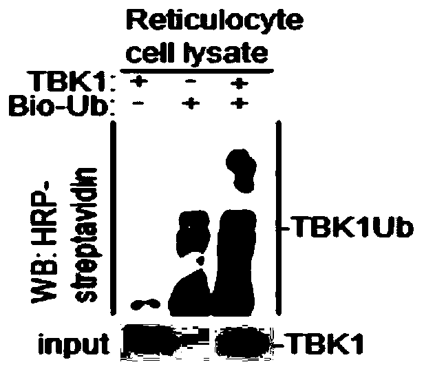
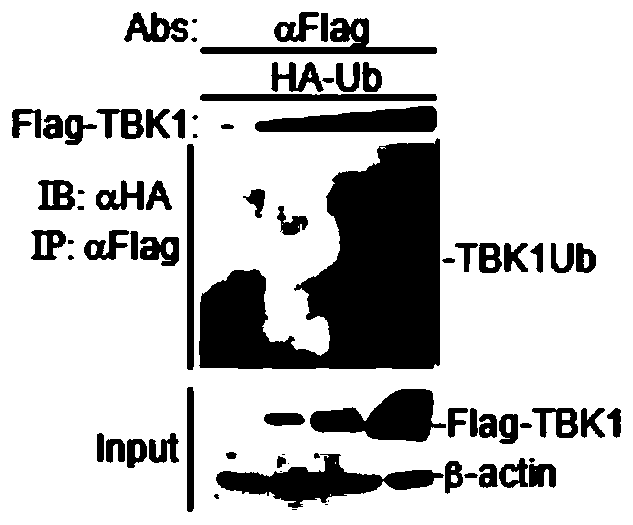
[0034] Example 1 Study on the activity of TBK1 as an E3 ubiquitin ligase

[0035] 1.1 In vitro ubiquitination experiment of TBK1

[0036] 1.1.1 In vitro transcription of plasmids

[0037] (1) According to the instructions of the in vitro transcription kit (TNT Quick Coupled Transcription / TranslationSystems kit), use the pCDNA3.1-Flag-TBK1 plasmid (1 μg) to be detected and the control plasmid pCDNA3.1 (1 μg) with TNT Quick Master Mix ( 24μL), Methionine (1.2μL), 0.5μg / μL plasmamid (2μL), H 2 O (2.8 μL) was mixed to prepare a reactant;

[0038] (2) Put the above reactant at 30° C. for 60-90 min to obtain the in vitro transcription product pCDNA3.1-Flag-TBK1 protein.

[0039] 1.1.2 Ubiquitination analysis experiments:

[0040] (1) According to the instructions of the in vitro ubiquitin kit (Enzo Life Sciences), the above in vitro transcription product pCDNA3.1-Flag-TBK1 protein (7.5 μL) was mixed with dH 2 O (14μL), 10×Ubiquitionylation Buffer (5μL), 100U / ml IPP (10μL), 50mM...

Embodiment 2

[0055] Example 2 Degradation of FMDV structural protein VP3 signaling pathway molecular analysis

[0056] 2.1 Experimental steps

[0057] (1) HEK-293T cells were paved in 12-well plates, and when the cells grew to 60%-80%, the plasmids of signaling pathway molecules (pCDNA3.1-HA-VISA, pCDNA3.1-HA-RIG-I, pCDNA3. 1-HA-IRF3-5D, pCDNA3.1-HA-IRF7, pCDNA3.1-HA-TBK1, pCDNA3.1-HA-MITA, pCDNA3.1-HA-RIG-I, pCDNA3.1-HA-MDA5) 1 μg of each, respectively co-transfected with 1 μg of FMDV VP3 plasmid into 293T cells;

[0058] (2) Collect the sample 24 hours after transfection, wash it 1-2 times with cooled PBS, add 100 μL of SDS loading buffer for lysis, and detect the expression of VP3 protein by WB method.

[0059] 2.2 Experimental results

[0060] The result is as image 3 As shown, only in the presence of TBK1 plasmid, VP3 protein is not expressed, therefore, only TBK1 in the above signaling pathway molecules can inhibit the expression of VP3 protein, indicating that TBK1 can degrade ...

Embodiment 3

[0061] Example 3 Analysis of TBK1 degrading FMDV structural protein VP3

[0062] 3.1 Experimental steps

[0063] (1) HEK-293T cells were plated in 12-well plates, and when the cells grew to 60%-80%, transfected with pCDNA3.1-Flag-VP3 plasmid and pCDNA3.1-HA-TBK1 plasmid, in which pCDNA3.1-Flag - VP3 plasmid 1 μg (constant amount), pCDNA3.1-HA-TBK1 plasmid 0.5 μg, 1 μg, 1.5 μg, 2 μg (increasing variable), or pCDNA3.1-HA-TBK1 plasmid 2 μg (constant amount), pCDNA3.1-Flag - VP3 plasmid 0.5 μg, 1 μg, 1.5 μg, 2 μg (increasing variable) for co-transfection;

[0064] (2) Collect the sample 24 hours after transfection, wash it 1-2 times with cold PBS, add 100 μL of SDS loading buffer to lyse, and detect the expression of pCDNA3.1-Flag-VP3 plasmid and pCDNA3.1-HA-TBK1 plasmid by WB .

[0065] 3.2 Experimental results

[0066] The result is as Figure 4 Shown: Whether the content of the pCDNA3.1-Flag-VP3 plasmid remains unchanged and the variable of the pCDNA3.1-HA-TBK1 plasmid inc...


PUM


Abstract

The invention belongs to the field of bio-medicine, in particular to a new application of TBK1 as E3 ubiquitin ligase. The TBK1 (TANK binding kinase 1) is found to have a ubiquitination function, cannot only undergo ubiquitination in vitro, but also undergo ubiquitination in vivo, and is a new E3 ubiquitin ligase; The invention further finds that the TBK1 can degrade structural protein VP3 of picornaviridae viruses including foot-and-mouth disease virus (FMDV), enterovirus (EV71), encephalomyocarditis virus (EMCV) and seneca virus (SVV), in particular can specifically degrade the structural protein VP3 of the FMDV, and can be used for preparing drugs related to degradation of the foot-and-mouth disease virus protein, preventing the foot-and-mouth disease virus protein from assembling related drugs, and preventing or treating foot-and-mouth disease virus infection related drugs.

Description

technical field [0001] The invention belongs to the field of biomedicine, and specifically relates to the application of TBK1 as an E3 ubiquitin ligase. Background technique [0002] A virus is a non-cellular form composed of a nucleic acid molecule (DNA or RNA) and protein. A protective shell wraps a piece of DNA or RNA, which can use the host's cellular system to replicate itself. Viruses can infect almost all living organisms with cellular structures and can cause a variety of diseases. [0003] Picornaviridae is a family consisting of the smallest group of RNA viruses, mainly including enterovirus, rhinovirus, cardiovirus and aphth virus. Among them, foot-and-mouth disease belongs to the genus Aphthus virus, which is an important disease that infects cloven-hoofed animals caused by foot-and-mouth disease virus. Foot-and-mouth disease virus (FMDV) is a single-stranded positive-sense RNA virus with a genome length of about 8200bp. . The FMDV open reading frame encodes a...

Claims


Application Information

Patent Timeline
Patent Type & Authority Patents(China)
IPC IPC(8): C12N9/00C12N15/52A61K38/53A61P31/14
CPCA61K38/00A61P31/14C12N9/93C12Y603/02019
Inventor 郑海学李丹杨文萍任静静茹毅郝荣增张克山田宏杨帆刘湘涛
Owner LANZHOU INST OF VETERINARY SCI CHINESE ACAD OF AGRI SCI
Why Eureka
Try Eureka