Medical image imaging method and device, computer equipment and storage medium

A technology of medical imaging and imaging methods, applied in the field of image processing, can solve the problems of side effects of patients, long scanning time of dynamic enhanced scanning, inability to clearly determine lesions and lesion boundaries, etc., and achieve the effect of fast imaging time

Active Publication Date: 2019-08-20
SHANGHAI UNITED IMAGING INTELLIGENT MEDICAL TECH CO LTD
View PDF10 Cites 33 Cited by
  • Summary
  • Abstract
  • Description
  • Claims
  • Application Information

AI Technical Summary

Problems solved by technology

[0003] However, for cancer patients whose lesions cannot be clearly judged, whose boundaries are unclear, or who have further needs, it is necessary not only to improve the detection rate of MR images for the lesion area, but also to show the lesion more clearly.
Traditionally, the location of the lesion is determined by performing dynamic enhanced scans on additional patients. However, dynamic enhanced scans not only take a long time to scan, but also require intravenous contrast agents to be injected in advance.
The contrast agent not only contains the paramagnetic substance gadolinium, but also the discharge rate is related to the metabolic capacity of the human body, which leads to certain side effects on patients

Method used


Image

  • Medical image imaging method and device, computer equipment and storage medium
  • Medical image imaging method and device, computer equipment and storage medium
  • Medical image imaging method and device, computer equipment and storage medium

Examples


Embodiment Construction

[0047] In order to make the purpose, technical solution and advantages of the present application clearer, the present application will be further described in detail below in conjunction with the accompanying drawings and embodiments. It should be understood that the specific embodiments described here are only used to explain the present application, and are not intended to limit the present application.

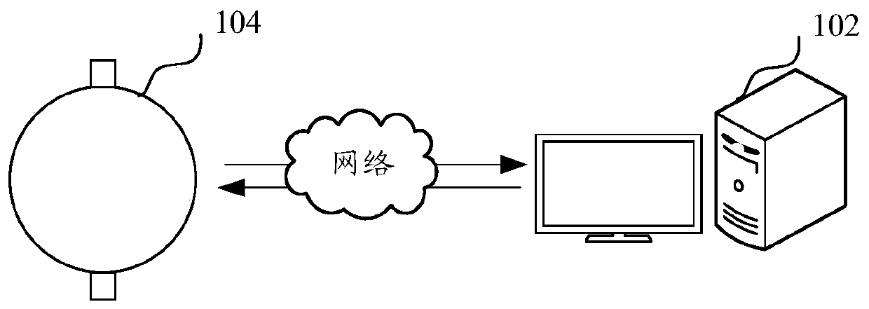
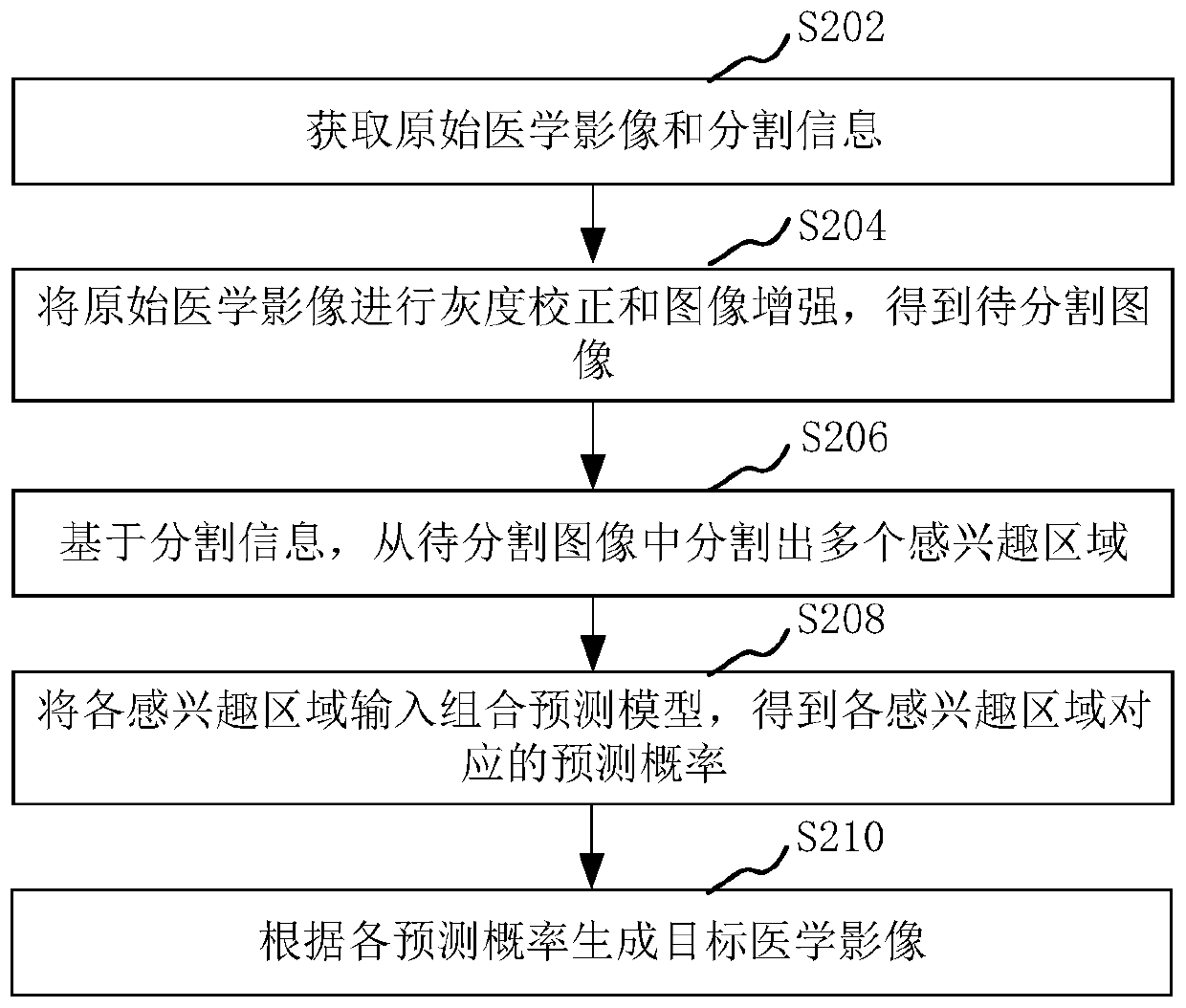
[0048] The medical imaging method provided by this application can be applied to such as figure 1 shown in the application environment. Wherein, the computer device 102 communicates with the medical imaging device 104 through a network. The computer device 102 obtains the original medical image and segmentation information, and the original medical image is obtained from the medical imaging device 104 . The computer device 102 performs grayscale correction and image enhancement on the original medical image to obtain the image to be segmented. Based on the segmentation ...


PUM


Abstract

The invention relates to a medical image imaging method and device, computer equipment and a storage medium. The method comprises the steps of obtaining an original medical image and segmentation information; performing gray correction and image enhancement on the original medical image to obtain a to-be-segmented image; segmenting a plurality of regions of interest from the to-be-segmented imagebased on the segmentation information; inputting each region of interest into the combined prediction model to obtain a prediction probability corresponding to each region of interest; and generatinga target medical image according to each prediction probability. By adopting the method, the time can be shortened, and no side effect is generated to patients.

Description

technical field [0001] The present application relates to the technical field of image processing, in particular to a medical image imaging method, device, computer equipment and storage medium. Background technique [0002] Magnetic resonance imaging (Magnetic Resonance Imaging, MRI) can know the position of atoms and draw the internal structure of objects through the magnetic field. Especially for the soft tissue layer display is very good. Therefore, MRI is commonly used as a diagnostic imaging tool for organ cancers, especially the prostate. Prostate is the most common type of cancer that threatens the lives of millions of men around the world, and is the second leading cause of death in men. MRI is an important diagnostic tool for prostate cancer imaging. MRI includes a variety of imaging modalities, such as T1-weighted imaging (T1W), T2-weighted imaging (T2W), diffusion-weighted imaging (DWI), apparent diffusion coefficient (ADC), and dynamic enhanced scan (DCE). -M...

Claims


Application Information

Patent Timeline
Patent Type & Authority Applications(China)
IPC IPC(8): G06T11/00G06T5/00G06T7/10
CPCG06T11/003G06T11/008G06T5/007G06T7/10G06T2207/10088G06T2207/20081G06T2207/20084
Inventor 徐小栋廖术詹翊强
Owner SHANGHAI UNITED IMAGING INTELLIGENT MEDICAL TECH CO LTD
Why Eureka
Try Eureka