Colorimetric method for detecting tobramycin based on double strand displacement and three-dimensional DNA structure

A technology for tobramycin and structure detection, applied in biochemical equipment and methods, microbiological determination/inspection, etc., can solve the problems of personnel training and sample preparation complexity, long test cycle, high detection limit, etc., and achieve the expansion of detection range , Improve detection sensitivity and specificity

Active Publication Date: 2019-12-20
JIANGNAN UNIV
View PDF3 Cites 10 Cited by
  • Summary
  • Abstract
  • Description
  • Claims
  • Application Information

AI Technical Summary

Problems solved by technology

However, these methods have some disadvantages, such as high detection limit, expensive equipment, long test cycle, personnel training and complicated sample preparation

Method used


Image

  • Colorimetric method for detecting tobramycin based on double strand displacement and three-dimensional DNA structure
  • Colorimetric method for detecting tobramycin based on double strand displacement and three-dimensional DNA structure
  • Colorimetric method for detecting tobramycin based on double strand displacement and three-dimensional DNA structure

Examples


Embodiment 1

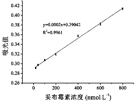
[0036] Example 1 Drawing of Tobramycin Concentration Standard Curve

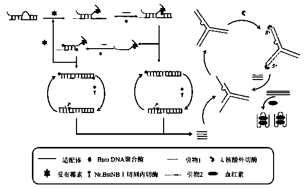
[0037] Mix T1 and T2 at the same concentration, denature at 95°C for 5 minutes, and then refold at 37°C for 120 minutes. 2 μL (10 μmol·L -1 ) double strands were mixed with 4 μL of tobramycin solutions of different concentrations, and incubated at 37°C for 30 min. Add 2 μL (10 μmol·L -1 ) Primer 1, 2 μL (10 μmol L -1 ) Primer 2, 5 U Nt.BstNBI nicking endonuclease, 8 U Bsm DNA polymerase and 2 μL (10 mmol·L -1 ) free deoxyribonucleoside triphosphate, 1× buffer (100 mmol·L -1 NaCl, 50mmol L -1 Tris-HCl, 10 mmol L -1 MgCl 2 , 0.1 mg·mL -1 BSA), mixed and incubated at 55°C for 120 min to generate a large amount of reporter probes, which were inactivated at 75°C for 10 min and stored at 4°C for use.

[0038] Mix 10 μL of S1, 10 μL of S2 and 10 μL of S3 (both 10 -6 mol L -1 ) were mixed, heated at 95 °C for 5 min, and then gradually cooled to room temperature. Thereafter, 30 μL of the DNA mixture ...

Embodiment 2

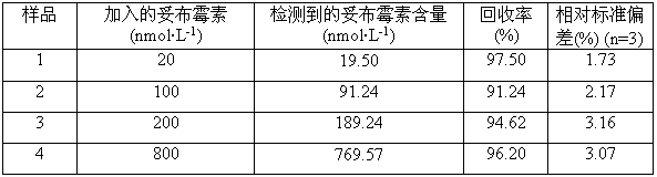
[0040] The determination of tobramycin content in the actual water sample of embodiment 2

[0041] In order to further verify the accuracy of this method in the determination of tobramycin content in actual samples, Taihu Lake water without pretreatment was selected to dilute tobramycin to different concentrations. The reaction was carried out in exactly the same way as the tobramycin standard sample, and the resulting reaction solution was read with a UV-vis spectrophotometer at 420nm absorbance, which was substituted into the standard curve to calculate the tobramycin concentration.

[0042] The specific samples and test results are shown in Table 1.

[0043] Table 1

[0044]


PUM


Abstract

The invention discloses a colorimetric method for detecting tobramycin based on double strand displacement and a three-dimensional DNA structure, and belongs to the field of food safety, medical analysis and environmental pollution detection. The method comprises the following steps: firstly, double strands T1 / T2 are designed; when tobramycin exists, Bsm DNA polymerase synthesizes double strands which are completely complementary through a strong strand displacement reaction, and Nt.BstNBI incision endonuclease cuts recognition sites on the double strands; the three-way DNA structure capture reporter probes, and regenerates and replaces a large number of S1 strands containing G-quadruplex forming sequences. Thereafter, the G-quadruplex / heme catalyzes ABTS<2-> / H2O2 chromogenic reaction, andthe tobramycin content can be determined by using the linear relationship between light absorption value and tobramycin concentration. According to the invention, an aptamer captures tobramycin to trigger double strand displacement reaction which is mediated by the Nt.BstNBI incision endonuclease and the Bsm DNA polymerase so as to generate a large number of reporter probes. Meanwhile, the reporter probes trigger lambda exonuclease-assisted loop amplification, so that multiple amplifications of colorimetric signals are realized, the detection range is widened, and the detection sensitivity isimproved.

Description

technical field [0001] The invention relates to a colorimetric method for detecting tobramycin based on double strand replacement and three-way DNA structure, and belongs to the fields of food safety, medical analysis and environmental pollution detection. Background technique [0002] Aminoglycoside antibiotics are antibiotics composed of two or more amino sugars linked to hexose rings through glycosidic bonds, and have good therapeutic effects on common bacterial diseases. Tobramycin is a broad-spectrum aminoglycoside antibiotic mainly used to treat infections caused by certain Gram-positive and aerobic Gram-negative microorganisms. Its mechanism is to bind to ribosomes, thereby destroying synthesized proteins, leading to cell membrane damage and cell death. Due to its good water solubility, low cost and broad antibacterial spectrum properties, tobramycin is widely used in animal husbandry. However, excessive and wrong use of tobramycin leads to large residues in food of...

Claims


Application Information

Patent Timeline
IPC IPC(8): C12Q1/682
CPCC12Q1/682C12Q2537/143C12Q2531/119C12Q2525/205C12Q2521/301
Inventor 周楠迪欧莹田亚平
Owner JIANGNAN UNIV
Why Eureka
Try Eureka