
Retina three-dimensional reconstruction method based on multiple fundus images without camera calibration

A fundus image and camera calibration technology, which is applied in 3D image processing, image data processing, 3D modeling, etc., can solve the problems of less research on 3D reconstruction of retinal surface area, complicated methods, and inability to be widely used. The reconstruction effect is good, the method is simple, and the effect of the retinal 3D reconstruction effect is good

Pending Publication Date: 2020-01-10
TIANJIN POLYTECHNIC UNIV
View PDF4 Cites 0 Cited by
  • Summary
  • Abstract
  • Description
  • Claims
  • Application Information

AI Technical Summary

Problems solved by technology

[0004] For the 3D reconstruction of fundus images, there have been preliminary studies in the literature. Most of the researches are on the local 3D reconstruction of the optic disc, and there are also studies on the 3D reconstruction of blood vessels, but there are few studies on the 3D reconstruction of the retinal surface area. Moreover, the camera needs to be calibrated, the method is complicated and cannot be widely used

Method used


Image

  • Retina three-dimensional reconstruction method based on multiple fundus images without camera calibration
  • Retina three-dimensional reconstruction method based on multiple fundus images without camera calibration
  • Retina three-dimensional reconstruction method based on multiple fundus images without camera calibration

Examples


Embodiment Construction

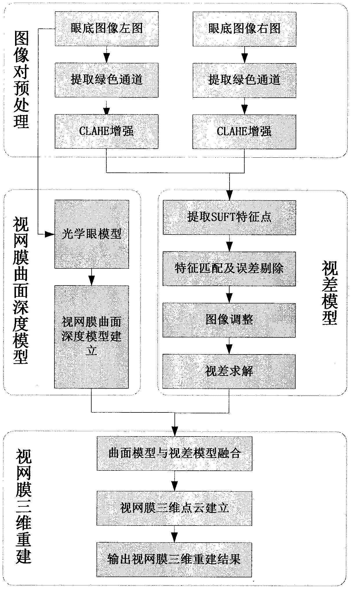
[0028] The flowchart of the present invention is as figure 1 As shown, the green channel of the fundus image pair is first extracted, and the contrast-limited adaptive histogram equalization algorithm is used to process the fundus image pair to enhance the contrast of the image. After that, the SUFT feature points are extracted, and the brute force matching method is used to initially match the feature points of the image pair. Then, the RANSAC algorithm and geometric constraint method are used to screen the initial matching feature points, and the affine transformation matrix between the image pairs is calculated, and the affine transformation matrix is ​​calculated according to the affine The projection transformation matrix adjusts the image, and finally the semi-global stereo matching algorithm (SGBM) is used to solve the parallax of the retina. Then, from the perspective of the design of the optical system of the fundus camera, a depth model of the retinal surface based on ...


PUM


Abstract

The invention discloses a retinal three-dimensional reconstruction method based on multiple fundus images without camera calibration. The method comprises the following steps: (1) collecting a fundusimage pair without calibrating a fundus camera, carrying out preprocessing and parallax solution on the fundus image, and establishing a retina surface parallax model; (2) establishing a retina curvedsurface depth model based on an optical eye model; and (3) fusing the retina curved surface depth model and the parallax model, establishing a retina three-dimensional point cloud, and completing three-dimensional reconstruction of the retina surface. When the method is adopted for fundus retina three-dimensional reconstruction, a fundus camera does not need to be calibrated, good robustness is achieved, and the three-dimensional reconstruction effect is good.

Description

Technical field [0001] The invention belongs to the field of image processing technology and fundus 3D reconstruction technology, and specifically relates to a method for 3D reconstruction of the retina based on multiple fundus images without camera calibration. The method does not require calibration of the fundus camera and has strong robustness. Most fundus images taken by fundus cameras are universally applicable. Background technique [0002] The retina of the human eye, also known as the fundus, is a layer of highly complex film that is an important part of the human eye. The position of the retina in the eye is equivalent to the receiver of the imaging system. The fundus is an important part of the sensory organ of the eye, and most of the diseases of the eye come from the fundus. At the same time, there are a large number of various arteriovenous blood vessels in the fundus, which can reflect the state of systemic vascular diseases. Diseases such as high blood pressure...

Claims


Application Information

Patent Timeline
Patent Type & Authority Applications(China)
IPC IPC(8): G06T15/00G06T17/00
CPCG06T15/005
Inventor 肖志涛耿磊娄世良张芳吴骏刘彦北王雯
Owner TIANJIN POLYTECHNIC UNIV
Learn More