Lung image segmentation method and device and lung lesion area identification equipment

An image segmentation and lung technology, applied in the field of image processing, can solve problems such as poor noise resistance, low seed point automation, and edge clutter

Inactive Publication Date: 2020-02-07
SHANGHAI MICROPORT PROPHECY MEDICAL TECH CO LTD
View PDF7 Cites 24 Cited by
  • Summary
  • Abstract
  • Description
  • Claims
  • Application Information

AI Technical Summary

Problems solved by technology

[0006] The essence of edge detection is to detect the edge according to the gray-scale jump gradient between neighboring pixels, and then perform segmentation and other processing. Considering the calculation method of the gradient, gradually introduce local differentials or gradients such as sobel, canny, roberts, and Gaussian operators. Operator, this method is more effective for edge detection, but often due to the existence of the gradient detection threshold, it may cause intermittent target boundaries, open boundaries, and edge clutter, and because it is sensitive to gradient information based on grayscale jumps, and The influence of noise cannot be eliminated, and the noise immunity is poor
[0007] The processing method based on region merging is to compare and classify pixels according to the similarity of pixels. There must be a starting point and benchmark for comparison, that is, seed points and merging growth rules. This method can merge pixels of the same nature in the same region, and then image It is divided into several different areas with different properties, which is simple and efficient, and it is clear at a glance. The disadvantage is that the degree of automation of the seed points is not high, and a certain amount of human intervention is required

Method used


Image

  • Lung image segmentation method and device and lung lesion area identification equipment
  • Lung image segmentation method and device and lung lesion area identification equipment
  • Lung image segmentation method and device and lung lesion area identification equipment

Examples


Embodiment Construction

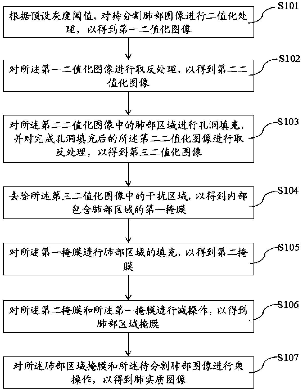
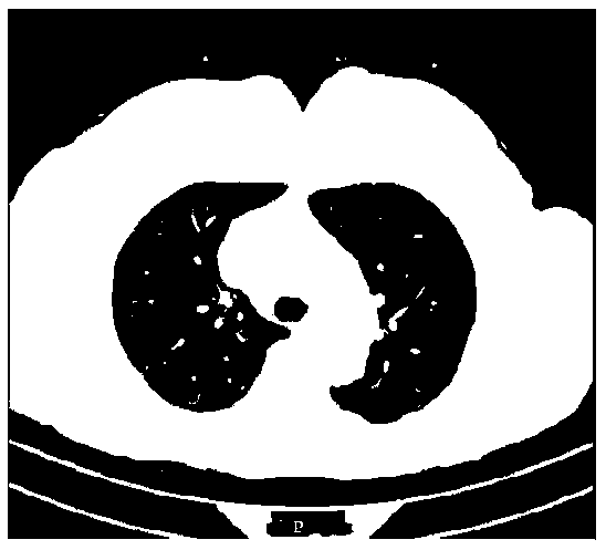
[0106] The following is attached Figures 1 to 7 and Specific Embodiments The lung image segmentation method, device, electronic equipment, storage medium and lung lesion area recognition equipment proposed in the present invention will be further described in detail. The advantages and features of the present invention will become clearer from the following description. It should be noted that the drawings are in a very simplified form and all use imprecise scales, which are only used to facilitate and clearly assist the purpose of illustrating the embodiments of the present invention. In order to make the objects, features and advantages of the present invention more comprehensible, please refer to the accompanying drawings. It should be noted that the structures, proportions, sizes, etc. shown in the drawings attached to this specification are only used to match the content disclosed in the specification, for those who are familiar with this technology to understand and re...


PUM


Abstract

The invention provides a lung image segmentation method and device and lung lesion area identification equipment, and the method comprises the steps: converting a to-be-segmented lung image into a first binary image according to a preset gray threshold; performing negation processing on the first binarized image to obtain a second binarized image; performing hole filling on the second binarized image, and performing negation processing to obtain a third binarized image; removing an interference region in the third binarized image to obtain a first mask; filling the first mask to obtain a second mask; performing subtraction operation on the second mask and the first mask to obtain a lung region mask; multiplying the lung region mask and the to-be-segmented lung image to obtain a lung parenchyma image. According to the invention, the lung parenchyma image can be extracted rapidly and accurately and lung lesion area identification is carried out by using the three-dimensional image, so that doctors are assisted well and the working efficiency is improved.

Description

technical field [0001] The present invention relates to the technical field of image processing, in particular to a lung image segmentation method, device, electronic equipment, storage medium, and lung lesion area recognition equipment. Background technique [0002] Chronic obstructive pulmonary disease (COPD), short for chronic obstructive pulmonary disease, is a common lung disease characterized by persistent respiratory symptoms and airflow limitation. According to incomplete statistics, more than 3 million people die from this disease every year in the world, and COPD has become the third leading cause of death. [0003] However, COPD is also a disease that can be prevented and diagnosed in advance. It has two major symptoms, namely chronic bronchitis and emphysema. Early lung CT images can be used to diagnose whether there is a lesion in the lung, so that doctors can further diagnose whether the patient has lung disease or whether there is a risk of disease. [0004]...

Claims


Application Information

Patent Timeline
Patent Type & Authority Applications(China)
IPC IPC(8): G06T7/136G06T7/187G06T5/40G06T5/00G06T5/30G06T17/00G06N3/04G06N3/08G06T7/00
CPCG06T7/136G06T7/187G06T5/40G06T5/002G06T5/30G06T17/00G06N3/08G06T7/0012G06T2207/10081G06T2207/30061G06N3/045
Inventor 张武龙吕文尔
Owner SHANGHAI MICROPORT PROPHECY MEDICAL TECH CO LTD
Why Eureka
Try Eureka