
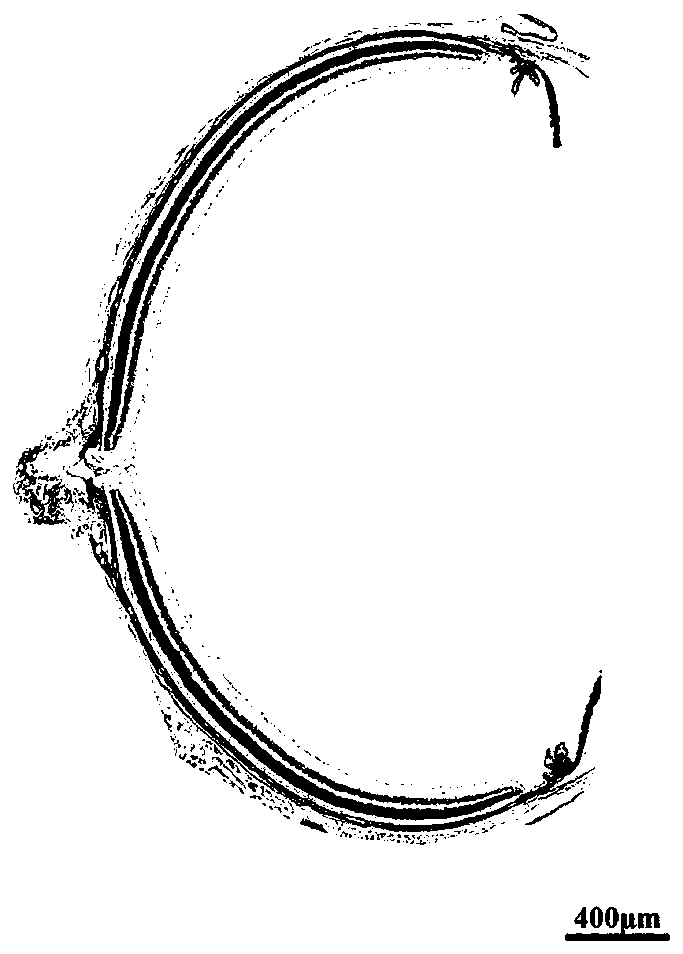
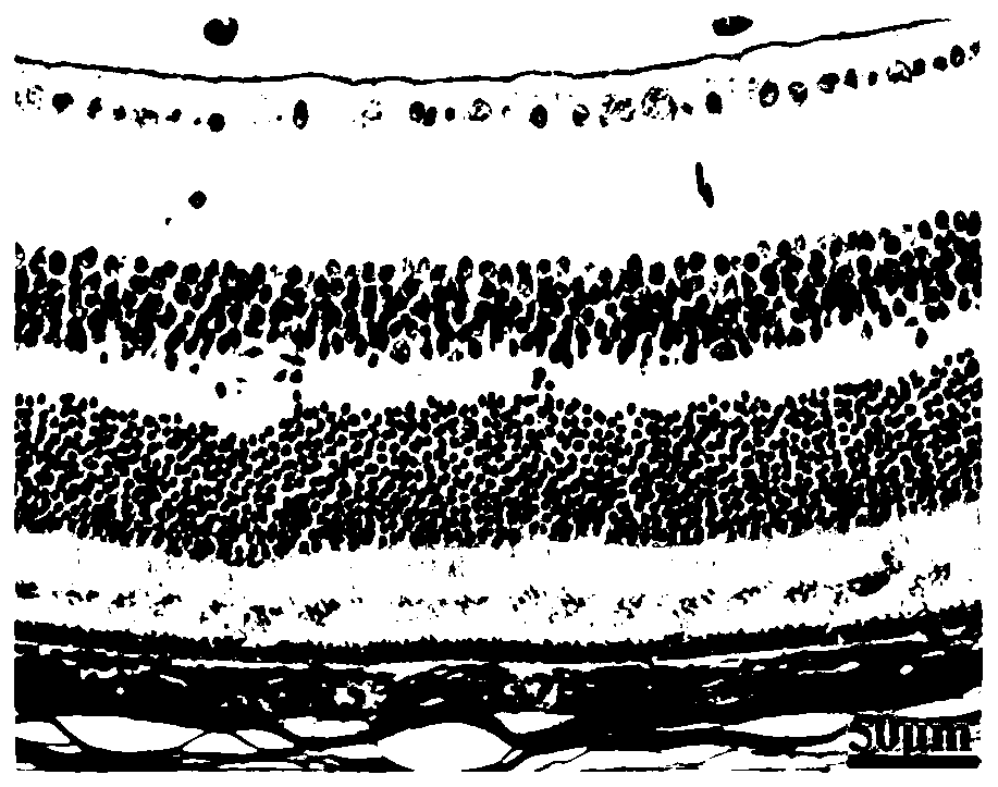
Preparation method of animal eyeball pathological section

A technology of pathological sections and production methods, applied in the field of histopathology, can solve problems such as eyeball deformation, detachment of various layers of eyeball, retinal tissue detachment, etc., to avoid detachment and improve quality.

Inactive Publication Date: 2020-02-21
THE FIFTH MEDICAL CENT OF CHINESE PLA GENERAL HOSPITAL
View PDF0 Cites 6 Cited by
  • Summary
  • Abstract
  • Description
  • Claims
  • Application Information

AI Technical Summary

Problems solved by technology

[0002] The pathological techniques routinely applied in the field of ophthalmology research involve the preparation of eyeball tissue specimens. At present, eyeball specimens fixed by conventional fixatives and conventional processing procedures are prone to deformation of the eyeball and detachment of various layers of the eyeball, especially retinal tissue. Detachment; low-quality pathological slices restrict high-quality pathological research in the field of ophthalmology; therefore, it is very necessary to develop a new type of fixative for animal eyeballs and optimize the use procedures to prepare high-quality eyeball pathological slices. Helps improve the basic application of retina-related research

Method used


Image

  • Preparation method of animal eyeball pathological section
  • Preparation method of animal eyeball pathological section
  • Preparation method of animal eyeball pathological section

Examples


Embodiment 1

[0032] The present embodiment provides a method for making a pathological section of an animal eyeball, comprising the following steps:

[0033] (1) Eyeball extraction: cut off the appendage muscles outside the eyeball of a small animal, and separate the eyeball; this embodiment uses the eyeball of a small animal as an example, and the small animal is an 8-week-old C57 mouse. Of course, other eyeballs with a thicker wall thickness can also be used Small animal eyeballs, such as rat eyeballs, etc.

[0034] (2) Eyeball fixation: put the isolated eyeball into a fixative solution pre-cooled to 4.0°C, and put it in a refrigerator set at 4.0°C for 48 hours; the composition and volume ratio of the fixative solution are as follows: 4 % paraformaldehyde solution: anhydrous acetic acid: absolute ethanol = 10: (1.5-2.0): (8.0-8.5).

[0035] (3) Dehydration and transparency of the optic cup of the eyeball: the eyeballs treated in (2) were treated with the following reagents in turn: 75% ...

Embodiment 2

[0040] The present embodiment provides a method for making a pathological section of an animal eyeball, comprising the following steps:

[0041] (1) Eyeball removal: cut off the extra-ocular appendage muscles of large animals, and separate the eyeballs; this embodiment uses the eyeballs of large animals as an example to illustrate, and the large animals are 24-week-old aborted fetuses. Of course, other eyeballs with thicker eyeball walls can also be used. Large animal eyeballs, such as rabbit eyeballs, cat eyeballs, pig eyeballs, monkey eyeballs, etc.

[0042] (2) Eyeball fixation: first use a needle to open several small holes in the central part of the cornea, for example, use a 33G needle to pierce 4 small holes in the central part of the cornea to accelerate the penetration of the fixative; then, put the separated eyeball into the Pre-cooled to 4.0°C in the fixative solution, put it in a refrigerator set at 4.0°C for 48 hours; the composition and volume ratio of the fixati...


PUM


Abstract

The invention relates to the field of histopathology, and particularly discloses a preparation method of an animal eyeball pathological section. The preparation method comprises the steps of: (1) eyeball extraction; (2) eyeball fixation, which is implemented by putting an separated eyeball into a stationary liquid which is pre-cooled to 4.0 DEG C, and putting the stationary liquid containing the eyeball into a refrigerator of which the temperature is set to 4.0 DEG C for fixation for 48 hours, wherein the stationary liquid comprises the following components: 4% paraformaldehyde solution, anhydrous acetic acid and absolute ethyl alcohol in a volume ratio of 10:(1.5-2.0):(8.0-8.5); (3) dehydration and transparent processing of an eyeball optic cup; (4) wax dipping and embedding; (5) and slicing. The preparation method is beneficial to preparation of the high-quality eyeball pathological section so as to facilitate improvement of basic application of retina related research.

Description

technical field [0001] The invention relates to the field of histopathology, in particular to a method for making pathological slices of animal eyeballs. Background technique [0002] The pathological techniques routinely applied in the field of ophthalmology research involve the preparation of eyeball tissue specimens. At present, eyeball specimens fixed by conventional fixatives and conventional processing procedures are prone to deformation of the eyeball and detachment of various layers of the eyeball, especially retinal tissue. Detachment; low-quality pathological slices restrict high-quality pathological research in the field of ophthalmology; therefore, it is very necessary to develop a new type of fixative for animal eyeballs and optimize the use procedures to prepare high-quality eyeball pathological slices. It helps to improve the basic application of retina-related research. Contents of the invention [0003] In view of this, the purpose of the present inventio...

Claims


Application Information

Patent Timeline
Patent Type & Authority Applications(China)
IPC IPC(8): G01N1/30G01N1/42G01N1/36G01N1/28
CPCG01N1/286G01N1/30G01N1/36G01N1/42G01N2001/2873G01N2001/305
Inventor 王少军杜璐杨艳峰黄慧敏
Owner THE FIFTH MEDICAL CENT OF CHINESE PLA GENERAL HOSPITAL
Learn More