Medical radiographic image phantom measurement standard device and phantom detection method

A measurement standard and phantom technology, applied in the field of medical measurement

Pending Publication Date: 2020-03-31
HENAN PROVINCE INST OF METROLOGY
View PDF4 Cites 3 Cited by
  • Summary
  • Abstract
  • Description
  • Claims
  • Application Information

AI Technical Summary

Problems solved by technology

[0004] The invention provides a medical radiographic phantom measurement standard device and a phantom detection method to solve the problem of traceability of the phantom, and provides an X-ray scanning three-dimensional tomographic imaging technology for obtaining the internal quality state and structural information of the medical radiographic phantom

Method used


Image

  • Medical radiographic image phantom measurement standard device and phantom detection method
  • Medical radiographic image phantom measurement standard device and phantom detection method
  • Medical radiographic image phantom measurement standard device and phantom detection method

Examples


Embodiment 1

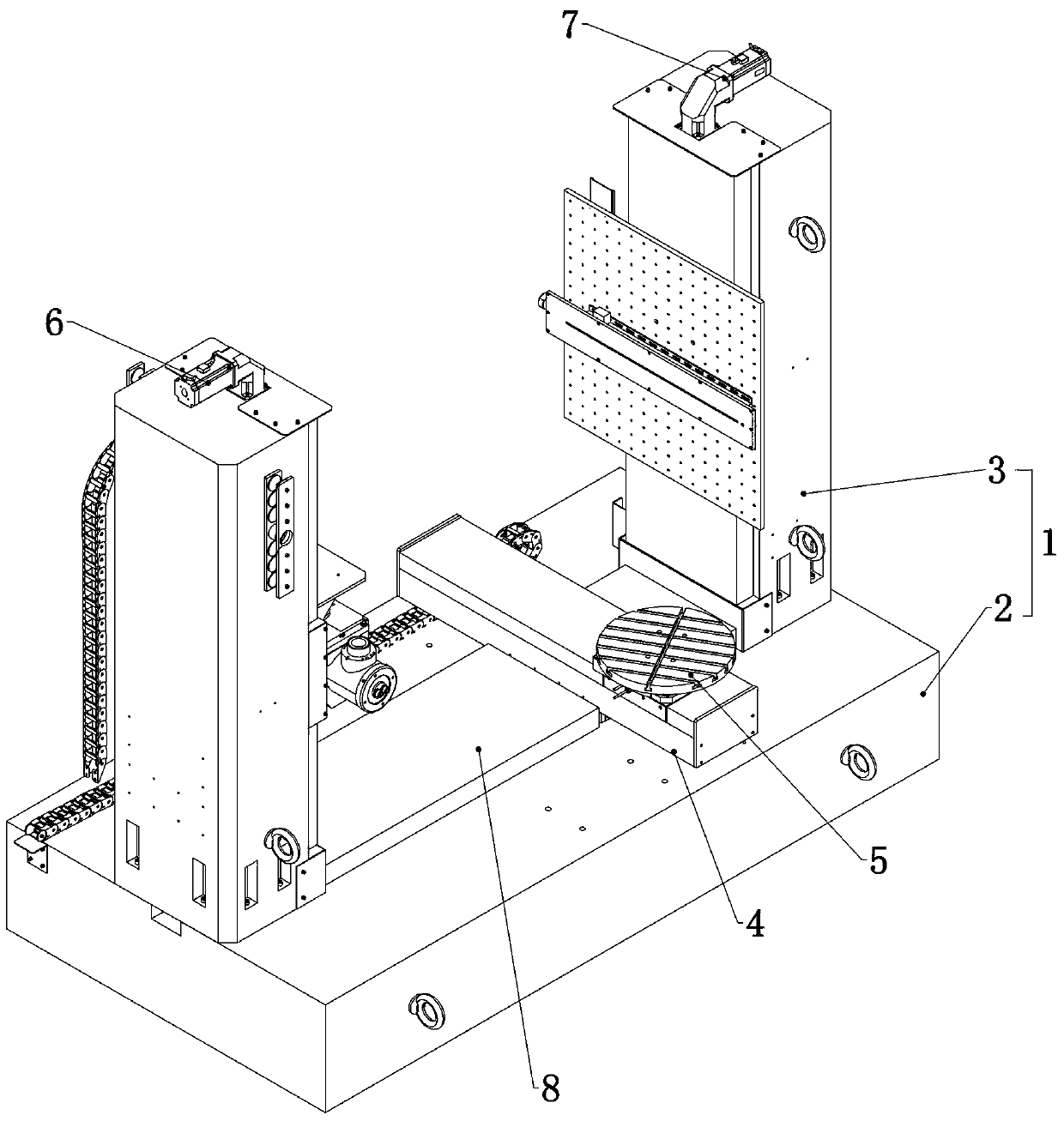
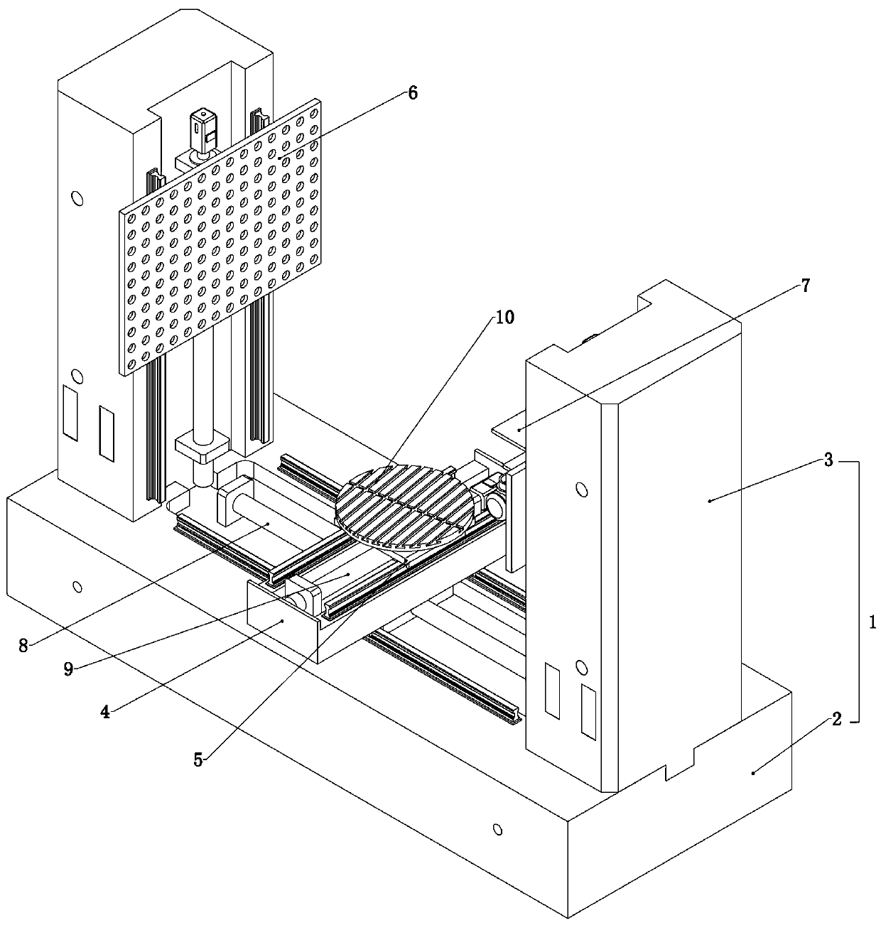
[0037] Embodiment 1, the present invention is a medical radiographic phantom measurement standard device, which is characterized in that it includes a platform 1, a motion mechanism, a ray tube 64 and a detector, and the platform 1 includes a base 2 and is relatively installed on the base 2 on the two uprights 3, refer to figure 1 , the base 2 is slidably connected with a workpiece motion platform 4, and the workpiece motion platform 4 is rotatably connected with a workpiece turntable 5, the workpiece turntable 5 is used to carry the device to be verified, and the ray tube 64 and detector Sliding up and down respectively connected to the opposite sides of the two columns 3;

[0038] The motion mechanism includes a ray tube vertical motion mechanism 6 and a detector vertical motion mechanism 7 which are respectively arranged on the opposite surfaces of the two columns 3, and the motion mechanism also includes a workpiece motion platform arranged on the base 2 transversely A mo...

Embodiment 2

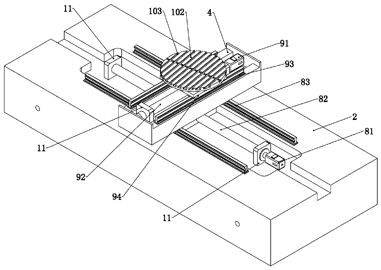
[0043] Embodiment 2. On the basis of Embodiment 1, the workpiece movement platform lateral motion mechanism 8 includes a lateral motor 81 fixedly connected in the base 2, and a lateral screw 82 is fixedly connected to the output shaft of the lateral motor 81. The transverse screw 82 passes through the workpiece motion platform 4 slidably connected to the base 2, and limit switches 11 are arranged at both ends of the transverse screw 82;

[0044] The base 2 is fixedly connected with a transverse slide rail 83, and the lower end of the workpiece moving platform 4 is fixedly connected with a transverse slide block 84 slidably connected to the transverse slide rail 83;

[0045] The vertical movement mechanism 9 of the turntable includes a vertical motor 91 fixedly connected to the workpiece moving platform 4, the output shaft of the vertical motor 91 is fixedly connected with a vertical screw 92, and the vertical screw 92 passes through The workpiece turntable 5 connected to the w...

Embodiment 3

[0051] Embodiment three, on the basis of embodiment two, also includes a control unit, a drive unit and a data acquisition image processing software system, the drive unit includes a servo motor drive module and a high-precision turntable drive module, and the servo motor drive The module is electrically connected to the horizontal motor 81, the vertical motor 91, the ray tube motor 61, and the detector motor 71, and the high-precision turntable drive module is electrically connected to the rotating motor 101;

[0052] The control unit is electrically connected to the drive unit, the detector, and the ray tube 64, and a data acquisition and image processing software system is installed inside;

[0053] It also includes radiation protection safety interlock and equipment protection safety interlock;

[0054] X-ray protection safety interlock includes X-ray machine door machine interlock, alarm light interlock and safety pedal or infrared interlock;

[0055] Equipment protectio...


PUM


Abstract

The invention relates to a medical radiographic image phantom measurement standard device and a phantom detection method, and provides an X-ray scanning three-dimensional tomography imaging device foracquiring the internal quality state and structural information of a medical radiographic image phantom. According to the technical scheme, the device comprises a platform, a movement mechanism, a ray tube and a detector, the platform comprises a base and two stand columns oppositely installed on the base, a workpiece movement platform is connected to the base in a sliding mode, a workpiece rotary table is connected to the workpiece movement platform in a rotating mode, and the ray tube and the detector are connected to the opposite faces of the two stand columns in an up-down sliding mode respectively. According to the invention, qualitative detection of material density is realized, and measurement of material CT value material density by industrial CT is realized.

Description

technical field [0001] The invention relates to the technical field of medical metrology, in particular to a medical radiographic phantom measurement standard device and a phantom detection method. Background technique [0002] Medical radiographic phantoms are used for metrological testing and quality control of radiological imaging equipment such as CT, CR / DR, DSA, etc. According to the national testing standards, regulations and specifications of CT, CR / DR, DSA and other medical radiological imaging equipment, it is necessary to use qualified phantoms for corresponding performance testing. At present, there is a lack of systematic evaluation methods for the performance evaluation of medical radiological imaging equipment in my country, and the problem of traceability of the measurement equipment of imaging equipment, that is, the phantom, has not been solved, which results in the lack of quality control of this type of equipment. [0003] Industrial Computed Tomography (...

Claims


Application Information

Patent Timeline
Patent Type & Authority Applications(China)
IPC IPC(8): G01N23/046G01N9/24
CPCG01N23/046G01N9/24G01N2223/03G01N2223/1016G01N2223/633
Inventor 王攀峰马长征王双玲袁晶晶徐凯李胜春姜鲲高申星龙成章刘辉唐盟高颖毛哲李静周云森王世闯张雪丽
Owner HENAN PROVINCE INST OF METROLOGY
Why Eureka
Try Eureka